Figure 2
Evidence of woven bone formation in carotid artery plaques
Mirzaie Masoud*, Zaur Guliyev, Michael Schultz, Peter Schwartz, Johann Philipp Addicks and Sheila Fatehpur
Published: 05 January, 2021 | Volume 6 - Issue 1 | Pages: 001-006
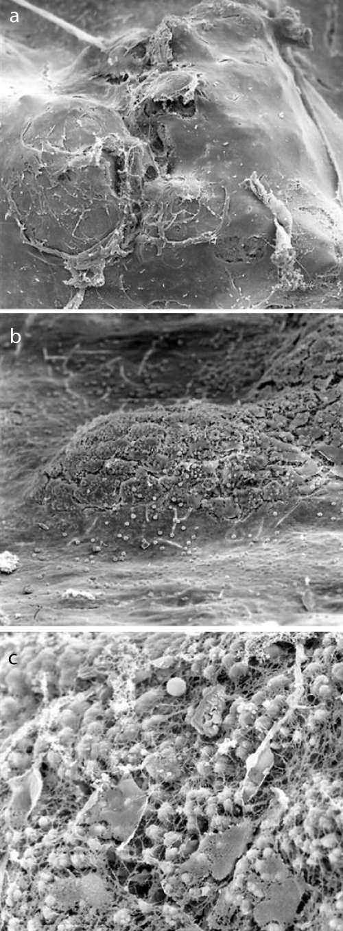
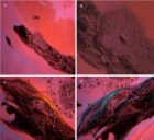
Figure 2:
a: Scanning electron microscopical findings of internal carotid plaque (50 x magnification): the surface of the observed plaques was uneven and partially broken. b: Scanning electron microscopical findings of internal carotid plaque (200 x magnification): multiple initial tearings and the uncovered extracellular matrix. c: Scanning electron microscopical findings of internal carotid plaque (1000 x magnification): incorporation of erythrocytes into the extracellular matrix with partially thin layer of endothelial cells.
Read Full Article HTML DOI: 10.29328/journal.jccm.1001108 Cite this Article Read Full Article PDF
More Images
Similar Articles
-
Evidence of woven bone formation in carotid artery plaquesMirzaie Masoud*,Zaur Guliyev,Michael Schultz,Peter Schwartz,Johann Philipp Addicks,Sheila Fatehpur. Evidence of woven bone formation in carotid artery plaques. . 2021 doi: 10.29328/journal.jccm.1001108; 6: 001-006
Recently Viewed
-
Assessment and Correlation of Serum Urea and Creatinine Levels in Normal, Hypertensive, and Diabetic Persons in Auchi, NigeriaAkpotaire PA, Seriki SA*. Assessment and Correlation of Serum Urea and Creatinine Levels in Normal, Hypertensive, and Diabetic Persons in Auchi, Nigeria. Arch Pathol Clin Res. 2023: doi: 10.29328/journal.apcr.1001035; 7: 007-016
-
Investigating Thermal Conductivity of FerrofluidsSumeir Walia*. Investigating Thermal Conductivity of Ferrofluids. Int J Phys Res Appl. 2023: doi: 10.29328/journal.ijpra.1001064; 6: 144-153
-
Cardiac Tamponade as the Cause of Pulmonary Edema: Case ReportEmídio Lima*. Cardiac Tamponade as the Cause of Pulmonary Edema: Case Report. J Pulmonol Respir Res. 2023: doi: 10.29328/journal.jprr.1001046; 7: 021-023
-
New Onset Seizures in a Child Taking 0.01% Atropine DropsCaitlyn Mulcahey*, Steve Gerber. New Onset Seizures in a Child Taking 0.01% Atropine Drops. Int J Clin Exp Ophthalmol. 2023: doi: 10.29328/journal.ijceo.1001051; 7: 003-005
-
Quality of Life (QoL) among Pakistani Women with Breast Cancer Undergoing ChemotherapyMohammad Yousaf*, Rita Ramos, Rehmatullah Inzar Gull. Quality of Life (QoL) among Pakistani Women with Breast Cancer Undergoing Chemotherapy. Arch Cancer Sci Ther. 2023: doi: 10.29328/journal.acst.1001037; 7: 018-026
Most Viewed
-
Evaluation of Biostimulants Based on Recovered Protein Hydrolysates from Animal By-products as Plant Growth EnhancersH Pérez-Aguilar*, M Lacruz-Asaro, F Arán-Ais. Evaluation of Biostimulants Based on Recovered Protein Hydrolysates from Animal By-products as Plant Growth Enhancers. J Plant Sci Phytopathol. 2023 doi: 10.29328/journal.jpsp.1001104; 7: 042-047
-
Feasibility study of magnetic sensing for detecting single-neuron action potentialsDenis Tonini,Kai Wu,Renata Saha,Jian-Ping Wang*. Feasibility study of magnetic sensing for detecting single-neuron action potentials. Ann Biomed Sci Eng. 2022 doi: 10.29328/journal.abse.1001018; 6: 019-029
-
Sinonasal Myxoma Extending into the Orbit in a 4-Year Old: A Case PresentationJulian A Purrinos*, Ramzi Younis. Sinonasal Myxoma Extending into the Orbit in a 4-Year Old: A Case Presentation. Arch Case Rep. 2024 doi: 10.29328/journal.acr.1001099; 8: 075-077
-
Pediatric Dysgerminoma: Unveiling a Rare Ovarian TumorFaten Limaiem*, Khalil Saffar, Ahmed Halouani. Pediatric Dysgerminoma: Unveiling a Rare Ovarian Tumor. Arch Case Rep. 2024 doi: 10.29328/journal.acr.1001087; 8: 010-013
-
Physical activity can change the physiological and psychological circumstances during COVID-19 pandemic: A narrative reviewKhashayar Maroufi*. Physical activity can change the physiological and psychological circumstances during COVID-19 pandemic: A narrative review. J Sports Med Ther. 2021 doi: 10.29328/journal.jsmt.1001051; 6: 001-007

HSPI: We're glad you're here. Please click "create a new Query" if you are a new visitor to our website and need further information from us.
If you are already a member of our network and need to keep track of any developments regarding a question you have already submitted, click "take me to my Query."


