More Information
Submitted: December 15, 2020 | Approved: February 11, 2021 | Published: February 12, 2021
How to cite this article: Ghitun FA, Ailoaei S, Ursu D, Chistol R, Tinica G. An unusual presentation of atrioventricular nodal reentrant tachycardia. J Cardiol Cardiovasc Med. 2021; 6: 014-018.
DOI: 10.29328/journal.jccm.1001110
Copyright License: © 2021 Ghitun FA, et al. This is an open access article distributed under the Creative Commons Attribution License, which permits unrestricted use, distribution, and reproduction in any medium, provided the original work is properly cited.
An unusual presentation of atrioventricular nodal reentrant tachycardia
Florina-Adriana Ghitun1, Stefan Ailoaei1, Dan Ursu2, Raluca Chistol3, Grigore Tinica4,5, Cristian Statescu1,4 and Mihaela Grecu1*
1Department of Cardiology, Institute for Cardiovascular Diseases, Iasi, Romania
2Department of Cardiology, Clinical Rehabilitation Hospital, Iasi, Romania
3Department of Radiology and Medical Imaging, Institute for Cardiovascular Diseases, Iasi, Romania
4Grigore T Popa University of Medicine and Pharmacy, Iasi, Romania
5Department of Cardiovascular Surgery, Institute for Cardiovascular Diseases, Iasi, Romania
*Address for Correspondence: Mihaela Grecu, MD, Institute for Cardiovascular Diseases, 50 Carol I Boulevard, Iasi, Romania, Tel: +40730650679; Email: mihaelagrecu@yahoo.com
Introduction: Atrioventricular nodal reentrant tachycardia (AVNRT) is the most frequent supraventricular tachycardia, commonly manifesting as autolimited paroxysmal episodes of rapid regular palpitations that exceed 150 beats per minute (bpm), dizziness and pounding neck sensation.
Case presentation: We present a case of a male patient, 70 years old, with ischemic heart disease and slow-fast AVNRT treated with radiofrequency catheter ablation (RFCA) in March 2019, with regular 6-months follow-ups. He was readmitted in our department in November 2020 for rest dyspnea and incessant fluttering sensation in the neck, without palpitations. The event electrocardiogram (ECG) was initially interpreted by general cardiologist as accelerated junctional rhythm, 75 bpm. Due to the persistence of symptoms and ECG findings, a differential diagnosis between reentry and focal automaticity was imposed. The response to vagal maneuvers and Holter ECG monitoring characteristics provided valuable information. We suspected recurrent slow ventricular rate typical AVNRT, which was confirmed by electrophysiological study and we successfully performed the RFCA of the slow intranodal pathway.
Conclusion: AV nodal reentry tachycardia may have an unusual presentation, occurring in elder male patients with structural heart disease. Antiarrhythmic drugs can promote reentry in this kind of patients. In cases of slow ventricular rate, vagal maneuvers and Holter ECG monitoring can help with the differential diagnosis. The arrhythmia can be successfully treated with RFCA with special caution regarding the risk of AV block.
Atrioventricular nodal reentrant tachycardia (AVNRT) is the most frequent supraventricular tachycardia (SVT) [1]. It is a paroxysmal, regular, narrow QRS tachycardia, with physiological substrate involving dual electrical pathways within the atrioventricular node (AV) [2-4]. There are two forms of AVNRT: typical (slow-fast) and atypical (fast-slow or slow-slow) [5]. Patients, which are usually female without structural heart disease, commonly present with auto-limited paroxysmal episodes of palpitations, dizziness and pounding neck sensation, with a ventricular rate that exceeds 150 beats per minute (bpm) [6-8].
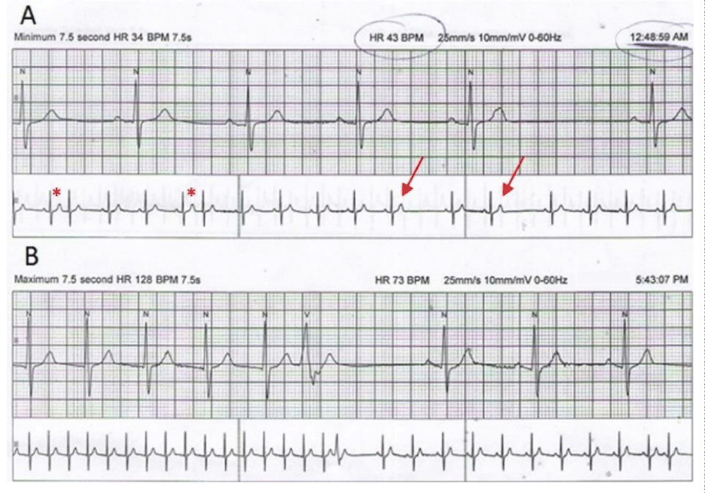
We present the case of a 70-year-old male patient, who came to our Arrhythmia outpatient clinic in June 2020 for rest dyspnea, incessant fluttering sensation in the neck and palpitations. His past medical history included a coronary artery bypass surgery (CABG) in 2012, first degree atrioventricular block (1stt degree AVB) and slow-fast AVNRT since 2016. Due to the frequent episodes of palpitations despite the beta-blocker treatment, he underwent a radiofrequency catheter ablation (RFCA) of the slow intranodal pathway in March 2019. He had no recurrence of the arrythmia at his regular 6 months follow-ups. He remained asymptomatic until April 2020, when the current symptomatology emerged, and he presented in another clinic. His electrocardiogram (ECG) was interpreted by the general cardiologist as accelerated junctional rhythm. He had a 24 hours Holter ECG monitoring, which documented sinus rhythm with an average heart rate of 70 bpm, but also 3 episodes of supraventricular tachycardia - 128 bpm, 7.5 seconds the longest, with sudden onset and termination (Figure 1) for which the cardiologist prescribed oral Amiodarone 400 mg.
Figure 1: Holter ECG monitoring strips: A. Minimum heart rate, 2 atrial extrasystoles with long PR interval (asterisks) and blocked P waves (arrows); B. Episode of supraventricular tachycardia, 128 bpm, showing sudden termination; 1 ventricular ectopic beat with VA conduction.
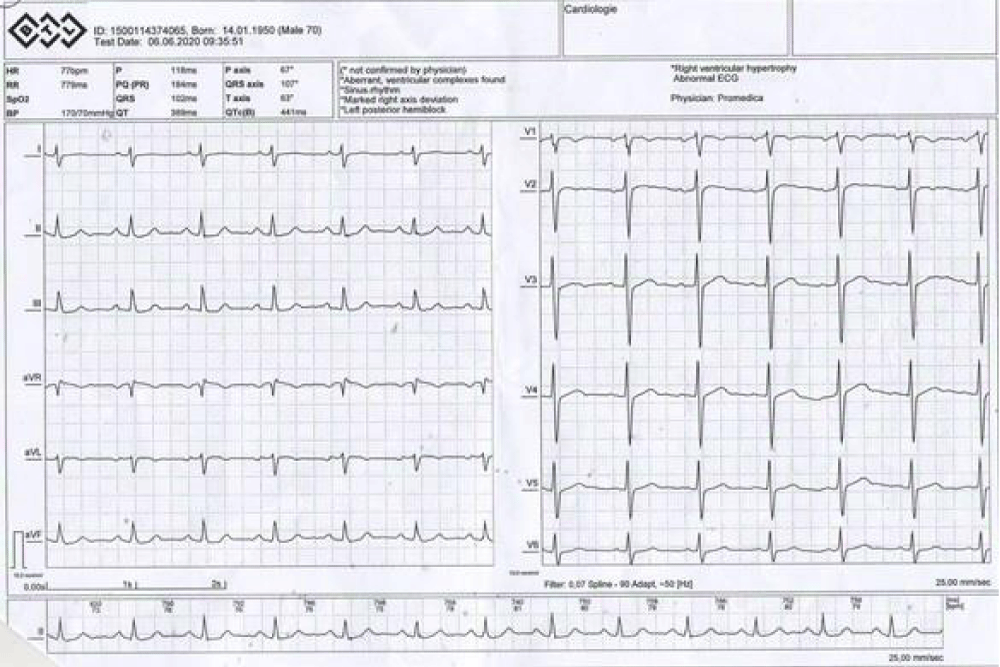
On presentation in our clinic, the physical examination showed intermittent neck pulsations, weak distal arterial pulse, postoperative scars and abdominal obesity (BMI = 30.9 kg/m2). The first ECG recorded showed sinus rhythm, 75 bpm, right axis deviation and a normal P wave and QRS morphology (Figure 2).
Figure 2: Basal ECG showing sinus rhythm.
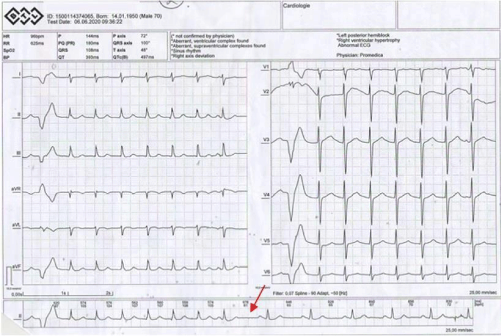
On a second ECG, we observed a supraventricular rhythm with narrow QRS complex, with retrograde P wave deforming the end of the QRS complex, 95 bpm. The arrhythmia was responsive to carotid sinus massage, but rapidly recurrent (Figure 3).
Figure 3: ECG during carotid sinus massage demonstrating transient conversion of supraventricular rhythm to sinus rhythm, suggestive for atrioventricular nodal reentry (arrow); 1 ectopic ventricular beat with VA conduction which initiates the arrhythmia.
Taking into consideration the clinical and electrophysiological features of the arrhythmia, we suspected the recurrence of the AVNR rather than an accelerated junctional rhythm. We withheld the Amiodarone treatment and the patient was scheduled for an electrophysiological study (EPS) in November 2020.
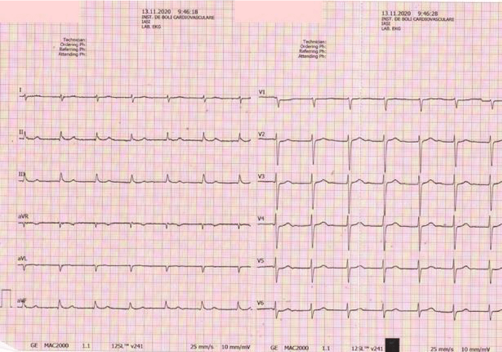
On admission, he declared the persistence of the symptomatology with neck pulsation and dyspnea. The ECG recorded showed the same supraventricular arrhythmia, 75/min (Figure 4). The blood tests did not reveal any electrolyte imbalance.
Figure 4: ECG at admission showing supraventricular arrhythmia.
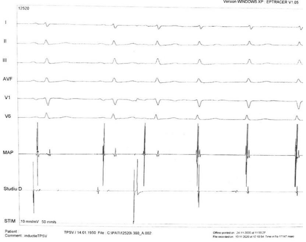
After acknowledging the potential risks and benefits, a written informed consent was obtained from the patient prior to the EPS. We used a right femoral approach. The basal measurements documented supra- and infrahisian conduction abnormalities, with atrial-His interval (AH) =123 ms and His-ventricle interval (HV) = 58 ms. Initially, we converted the arrhythmia to sinus rhythm by overdrive stimulation. The arrhythmia was induced again both spontaneously and with a single atrial extrastimuli – hence suggesting a reentry mechanism, with sudden prolongation of the AH interval with 90 ms and a VA interval of 0ms in the area adjacent to the coronary sinus ostium (Figure 5). We performed an apical right ventricular stimulation during the arrhythmia and we obtained a V-A-V response which excluded the diagnosis of atrial tachycardia. Concentrical and decremental VA conduction excluded the presence of an occult accesory pathway. Therefore, we made the diagnosis of slow-fast AVNR with the cycle length of 800ms (75 bpm).
Figure 5: Atrioventricular nodal reentry induction with an atrial extrastimuli coupled at 300 ms, demonstrating AH jump of 90 ms and superimposed V and A (VA interval = 0 ms) in the periosteal area.
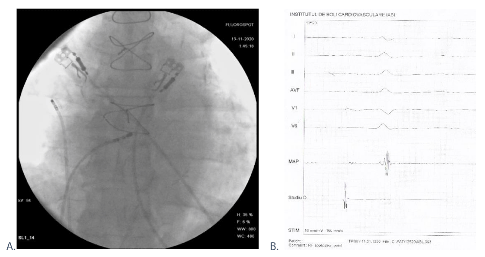
After identifying the Koch triangle elements, we localized the slow intranodal pathway anatomically – in a posterior-anterior radiological position (Figure 6A) and electrophysiologically – where the atrial deflection was small, with an atrium to ventricle ratio of 1 to 10. At this site, we performed 0:55 minutes (min) of radiofrequency applications (Figure 6B). For safety reasons, we did not reach maximal parameters of power and temperature and we frequently checked the ablation catheter position fluoroscopically, considering his AV conduction abnormalities. Post-ablation, the atrioventricular conducting parameters remained unchanged. We verify the non-inducibility of the arrhythmia using the atrial stimulation protocol by introducing up to 6 atrial extrastimuli until the nodal atrioventricular refractoriness was achieved, in basal conditions and after 1 mg of Atropine. The exposure time was 6:16 min with a radiation dose of 4995 μGy/m2 and the total procedure time was 80 min.
Figure 6: Ablation site: A. Fluoroscopy, posterior-anterior position, showing the placement of the study catheter on the lateral wall of right atrium and ablation catheter within the Koch triangle. B. Electrogram during ablation, A to V ratio = 1:10.
The post-procedural evolution was uneventful, without recurrence of the arrhythmia clinically or on ECG. We discharged the patient the next day, maintaining the beta-blocker for its anti-ischemic properties. One month post-ablation, the patient remained asymptomatic.
Our case presents a series of particularities which we would like to highlight:
1. The presentation of the patient was atypical. AVNRT is more common in young adults, without structural heart disease, and women are more likely to be affected than men (ratio 70:30) [6-8]. In addition, patients more commonly report paroxysmal episodes of regular tachycardia with sudden onset, palpitations being the most frequent symptom declared (98% of patients), alongside dizziness, dyspnea and neck pulsation1. Although our patient presented some of these characteristics, he was male, old, with ischemic heart disease, he did not report palpitations and the character of his arrhythmia was incessant. A possible explanation for the last two features is his relatively slow ventricular rate of 75 bpm, which can promote the re-entry mechanism, making it almost permanent and is within the normal sinus range, hence not causing palpitations. A ventricular rate less than 100 bpm is very rare in AVNRT [7,9]. Previous studies associated a slow rate AVNRT with advanced age, higher Wenckebach cycle length, AV node antegrade effective refractory period values and higher atrial vulnerability [10-13]. In our patient this could be associated with his AV conduction abnormalities, the previous RFCA procedure and the beta-blocking and Amiodarone treatment [14].
2. The event ECG was not characteristic for typical (or atypical) AVNRT and a differential diagnosis with accelerated junctional rhythm/junctional ectopic tachycardia was necessary. The rate was in the range of 60 to 130 bpm, with the P waves deforming the end of the QRS complex [15] and the patient had ischemic heart disease with CABG surgery. Nevertheless, these medical antecedents were remote from the presentation moment, there was no sick sinus syndrome which would have justified an escape junctional rhythm and we identified a series of electrophysiological features that helped us suspecting the diagnosis of slow ventricular rate typical AVNRT:
• The arrhythmia was responsive to vagal maneuvers, carotid sinus massage suddenly terminating the tachycardia [16-19].
• On Holter ECG monitoring there were episodes of supraventricular tachycardia (> 120 bpm), with sudden onset and termination; also, we could identify single premature atrial beats with PR prolongation, thus suggesting a dual nodal physiology [20].
All these findings were in favor of a reentry mechanism rather than a focal automaticity and we confirmed the diagnosis during the EPS, by inducing and terminating the arrhythmia with single atrial extrastimulation [20,21].
3. In both ESC and ACC/AHA supraventricular tachycardias guidelines, the RFCA of the slow intranodal pathway is a first-line therapy (class IB recommendation) in symptomatic patients due to its high rate of success (> 95%) and low risk profile [9,22]. In our case, we consider successful the first procedure because the patient remained asymptomatic for 12 months. The recurrence was probably a consequence of the submaximal parameters (power, temperature) of the radiofrequency applications imposed by the AV conduction disturbance, in order to avoid an AV block [10,23].
Although AVNRT is more prevalent in young healthy women and it has established ECG diagnostic criteria, it may also occur in elder male patients, with structural heart disease. Furthermore, a heart rate less than 100 bpm does not exclude the diagnosis. In these particular situations, ECG recordings during vagal maneuvers and Holter ECG monitoring can help with the differential diagnosis, which can be further confirmed and treated invasively trough EPS and RFCA.
- Katritsis DG, Camm AJ. Classification and differential diagnosis of atrioventricular nodal re-entrant tachycardia. Europace. 2006; 8: 29–36. PubMed: https://pubmed.ncbi.nlm.nih.gov/16627405/
- Nikolaidou T, Aslanidi OV, Zhang H, Efimov IR. Structure-function relationship in the sinus and atrioventricular nodes. Pediatr Cardiol. 2012; 33: 890-899. PubMed: https://pubmed.ncbi.nlm.nih.gov/22391764/
- Hucker WJ, McCain ML, Laughner JI, Iaizzo PA, Efimov IR. Connexin 43 expression delineates two discrete pathways in the human atrioventricular junction. Anat Rec (Hoboken). 2008; 291: 204-215. PubMed: https://pubmed.ncbi.nlm.nih.gov/18085635/
- Katritsis DG, Efimov IR. Cardiac connexin genotyping for identification of the circuit of atrioventricular nodal re-entrant tachycardia. Europace. 2018; 21: 190-191. PubMed: https://pubmed.ncbi.nlm.nih.gov/29860485/
- Katritsis DG, Camm AJ. Atrioventricular nodal reentrant tachycardia. Circulation. 2010; 122: 831-840. PubMed: https://pubmed.ncbi.nlm.nih.gov/20733110/
- Porter MJ, Morton JB, Denman R, Lin AC, et al. Influence of age and gender on the mechanism of supraventricular tachycardia. Heart Rhythm. 2004; 1: 393–396. PubMed: https://pubmed.ncbi.nlm.nih.gov/15851189/
- Gonzalez-Torrecilla E, Almendral J, Arenal A, Atienza F, Atea LF, et al. Combined evaluation of bedside clinical variables and the electrocardiogram for the differential diagnosis of paroxysmal atrioventricular reciprocating tachycardias in patients without pre-excitation. J Am Coll Cardiol. 2009; 53:2353–2358. PubMed: https://pubmed.ncbi.nlm.nih.gov/19539146/
- Liuba I, Jönsson A, Säfström K, Walfridsson H. Gender-related differences in patients with atrioventricular nodal reentry tachycardia. Am J Cardiol. 2006; 97: 384-388. PubMed: https://pubmed.ncbi.nlm.nih.gov/16442401/
- Page RL, Joglar JA, Caldwell MA, Calkins H. Conti JB, et al. Evidence Review Committee Chair‡. 2015 ACC/AHA/HRS Guideline for the Management of Adult Patients with Supraventricular Tachycardia: Executive Summary: A Report of the American College of Cardiology/American Heart Association Task Force on Clinical Practice Guidelines and the Heart Rhythm Society. Circulation. 2016; 133: e471-505. PubMed: https://pubmed.ncbi.nlm.nih.gov/26399662/
- Grecu M, Floria M, Arsenescu-Georgescu C. Abnormal Atrioventricular Node Conduction and Atrioventricular Nodal Reentrant Tachycardia in Patients Older Versus Younger Than 65 Years of Age. Pacing Clin Electrophysiol. 2009; 32: S98-100. PubMed: https://pubmed.ncbi.nlm.nih.gov/19250123/
- Pentinga ML, Meeder JG, Crijns HJ, de Muinck GM, Wiesfeld ED, et al. Late onset atrioventricular nodal tachycardia. Int J Cardiol. 1993; 38: 293-298. PubMed: https://pubmed.ncbi.nlm.nih.gov/8463010/
- Evrengul H, Alihanoglu YI, Kilic I, Yildiz S, Kose S. Clinical and electrophysiological characteristics of the patients with relatively slow atrioventricular nodal reentrant tachycardia. J Interv Card Electrophysiol. 2014; 40: 117–123. PubMed: https://pubmed.ncbi.nlm.nih.gov/24793102/
- Vijayaraman P, Alaeddini J, Storm R, Oren J, Wood MA, et al. Slow Atrioventricular Nodal Reentrant Arrhythmias: Clinical Recognition, Electrophysiological Characteristics, and Response to Radiofrequency Ablation. Journal of Cardiovascular Electrophysiology 2007; 18: 950-953. https://pubmed.ncbi.nlm.nih.gov/17666062/
- Wellens HJJ, Brugada P, Abdollah H. Effect of amiodarone in paroxysmal supraventricular tachycardia with or without Wolff-Parkinson-White syndrome. Am Heart J. 1983; 106: 876-880. PubMed: https://pubmed.ncbi.nlm.nih.gov/6613833/
- Katritsis DG, Josephson ME. Differential diagnosis of regular, narrow-QRS tachycardias. Heart Rhythm. 2015; 12: 1667-1676. PubMed: https://pubmed.ncbi.nlm.nih.gov/25828600/
- Smith GD, Fry MM, Taylor D, Morgans A, Cantwell K. Effectiveness of the Valsalva Manoeuvre for reversion of supraventricular tachycardia. Cochrane Database Syst Rev. 2015; 2:CD009502. PubMed: https://pubmed.ncbi.nlm.nih.gov/25922864/
- Lim SH, Anantharaman V, Teo WS, Goh PP, Tan ATH. Comparison of treatment of supraventricular tachycardia by Valsalva maneuver and carotid sinus massage. Ann Emerg Med. 1998; 31: 30-35. PubMed: https://pubmed.ncbi.nlm.nih.gov/9437338/
- Smith G, Morgans A, Boyle M. Use of the Valsalva manoeuvre in the prehospital setting: a review of the literature. Emerg Med J. 2009; 26: 8-10. PubMed: https://pubmed.ncbi.nlm.nih.gov/19104086/
- Green M, Heddle B, Dassen W, Wehr M, Abdollah H, et al. Value of QRS alteration in determining the site of origin of narrow QRS supraventricular tachycardia. Circulation. 1983; 68: 368-373.
- Silva RMF, Roever L. Typical atrioventricular nodal reentrant and orthodromic atrioventricular tachycardias: electrocardiographic, electrophysiological diagnosis and treatment. Interventional Cardiol. 2016; 8: 621-628.
- Glover BM, Brugada P. AV Nodal Re-entry Tachycardia (AVNRT)”, BM Glover & P Brugada, Springer International Publishing, Switzerland, Clinical Handbook of Cardiac Electrophysiology. 2016; 119-132.
- Brugada J, Katritsis DG, Arbelo E, Arribas F, Bax JJ, et al. 2019 ESC Guidelines for the management of patients with supraventricular tachycardia The Task Force for the management of patients with supraventricular tachycardia of the European Society of Cardiology (ESC). Eur Heart J. 2020; 41: 655-720. PubMed: https://pubmed.ncbi.nlm.nih.gov/31504425/
- Li YG, Grönefeld G, Bender B, Machura C, Hohnloser SH. Risk of development of delayed atrioventricular block after slow pathway modification in patients with atrioventricular nodal reentrant tachycardia and a pre-existing prolonged PR interval. Eur Heart J. 2001; 22: 89–95. PubMed: https://pubmed.ncbi.nlm.nih.gov/11133214/
