Abstract
Research Article
RV Function by cardiac magnetic resonance and its relationship to RV longitudinal strain and neutrophil/lymphocyte ratio in patients with acute inferior ST-segment elevation myocardial infarction undergoing primary percutaneous intervention
Salma Taha*, Shrouk Kelany Ali, Fabrizio D’Ascenzo, Hosam Hasan-Ali, Yousra Ghzally# and Mohamed Abdel Ghany
Published: 23 November, 2021 | Volume 6 - Issue 3 | Pages: 059-065
Background: Although acute inferior myocardial infarction (MI) is usually regarded as being lower risk compared with acute anterior MI, right ventricular (RV) myocardial involvement (RVMI) may show an increased risk of cardiovascular (CV) morbidity and mortality in patients with inferior MI. CMR is ideal for assessing the RV because it allows comprehensive evaluation of cardiovascular morphology and physiology without most limitations that hinder alternative imaging modalities.
Objectives: To evaluate the sensitivity of strain and strain rate of the RV using 2D speckle tracking echo and the neutrophil/ lymphocyte ratio (NLR) compared to cardiac MRI (CMR) as the gold standard among patients with inferior STEMI undergoing primary percutaneous coronary intervention (PCI).
Methodology: 40 Patients with inferior MI who had primary PCI were included in the study; they were divided into two groups according to the RVEF using CMR. NLR was done in comparison to RVEF.
Results: out of the 40 patients, 18 (45%) patients had RV dysfunction. 2D echocardiography was done for all patients, where fractional area change (FAC) in the RV dysfunction group appeared to be significantly reduced compared to the group without RV dysfunction (p value = 0.03). In addition, RV longitudinal strain (LS) by speckle tracking echo was reduced with an average of 19.5 ± 3.9% in the RV dysfunction group.
Both CMR- derived RV SV, and EF were lower among the RV dysfunction group, (26.8 ± 15.8) ml and (35.4 ± 6.9)% respectively, with large RV systolic volume, with a highly statistically significant difference in comparison to the other group (p value = 0.000). Complications, heart block was significantly higher in patients with RV dysfunction (p value = 0.008) as it occurred in 5 (27.8%) patients.
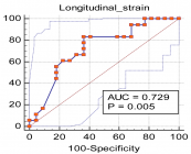
N/L ratio for predicting RV dysfunction by CMR had a cut-off value of > 7.7 with low sensitivity (38.8%) and high specificity (77.3 %). In contrast, LS for predicting RV dysfunction by CMR had high sensitivity (83.3%) and high specificity (63.6%) with p value = 0.005.
Conclusion: Our results showed that RV dysfunction in inferior MI is better detected using cardiac magnetic resonance imaging. In inferior STEMI patients who underwent primary PCI, NLR has low sensitivity but high specificity for predicting RVD when measured by cardiac MRI.
Read Full Article HTML DOI: 10.29328/journal.jccm.1001120 Cite this Article Read Full Article PDF
Keywords:
Right ventricular longitudinal strain; Neutrophil/lymphocyte ratio; Inferior wall myo-cardial infarction; Right ventricular dysfunction; Primary percutaneous coronary intervention; Cardiac magnetic resonance imaging; Right ventricular ejection fraction; Fractional area change
References
- Wang JY, Goodman SG, Saltzman I, Wong GC, Huynh T, et al. Cardiovascular risk factors and in-hospital mortality in acute coronary syndromes: insights from the Canadian global registry of acute coronary events. Can J Cardiol. 2015; 31: 1455-1461. PubMed: https://pubmed.ncbi.nlm.nih.gov/26143140/
- Goldstein JA. Pathophysiology and management of right heart ischemia. J Am College Cardiol. 2002; 40: 841-853. PubMed: https://pubmed.ncbi.nlm.nih.gov/12225706/
- Mehta SR, Eikelboom JW, Natarajan MK, Diaz R, Yi C, Gibbons RJ, et al. Impact of right ventricular involvement on mortality and morbidity in patients with inferior myocardial infarction. J A Coll Cardiol. 2001; 37: 37-43. PubMed: https://pubmed.ncbi.nlm.nih.gov/11153770/
- Ali ER, Mohamad AM. Diagnostic accuracy of cardiovascular magnetic resonance imaging for assessment of right ventricular morphology and function in pulmonary artery hypertension. Egyptian J Chest Dis Tuberculosis. 2017; 66: 477-4786.
- Yeh DD, Foster E. Is MRI the preferred method for evaluating right ventricular size and function in patients with congenital heart disease? MRI is not the preferred method for evaluating right ventricular size and function in patients with congenital heart disease. Circ Cardiovasc Imaging. 2014; 7: 198-205. PubMed: https://pubmed.ncbi.nlm.nih.gov/24449549/
- Wu WC, Rathore SS, Wang Y, Radford MJ, Krumholz HM. Blood transfusion in elderly patients with acute myocardial infarction. New Engl J Med. 2001; 345: 1230-1236. PubMed: https://pubmed.ncbi.nlm.nih.gov/11680442/
- Lang RM, Badano LP, Mor-Avi V, Afilalo J, Armstrong A, et al. Recommendations for cardiac chamber quantification by echocardiography in adults: an update from the American Society of Echocardiography and the European Association of Cardiovascular Imaging. J Am Soc Echocardiogr. 2015; 28: 001-039. PubMed: https://pubmed.ncbi.nlm.nih.gov/25559473/
- Frangogiannis NG. The immune system and cardiac repair. Pharmacol Res. 2008; 58: 88-111. PubMed: https://pubmed.ncbi.nlm.nih.gov/18620057/
- Thygesen K. Alpert JS, Jaffe AS, Simoons ML, Chaitman BR, et al. Third universal definition of myocardial infarction. Eur Heart J. 2012; 33: 2551-2567. PubMed: https://pubmed.ncbi.nlm.nih.gov/22922414/
- Ibánez B, James S, Agewall S, Antunes MJ, Bucciarelli-Ducci C, et al. 2017 ESC Guidelines for the management of acute myocardial infarction in patients presenting with ST-segment elevation. Eur Heart J. 2018; 39:119-177. PubMed: https://pubmed.ncbi.nlm.nih.gov/28886621/
- Hudsmith LE, Petersen SE, Francis JM, Robson MD, Neubauer S. Normal human left and right ventricular and left atrial dimensions using steady state free precession magnetic resonance imaging. J Cardiovasc Magn Reson. 2005; 7: 775-782. PubMed: https://pubmed.ncbi.nlm.nih.gov/16353438/
- Maceira AM, Prasad SK, Khan M, Pennell DJ. Reference right ventricular systolic and diastolic function normalized to age, gender and body surface area from steady-state free precession cardiovascular magnetic resonance. Eur Heart J. 2006; 27: 2879-2888. PubMed: https://pubmed.ncbi.nlm.nih.gov/17088316/
- Kakouros N, Cokkinos DV. Right ventricular myocardial infarction: pathophysiology, diagnosis, and management. Postgrad Med J. 2010; 86: 719-728. PubMed: https://pubmed.ncbi.nlm.nih.gov/20956396/
- Galea N, Francone M, Carbone I, Cannata D, Vullo F, et al. Utility of cardiac magnetic resonance (CMR) in the evaluation of right ventricular (RV) involvement in patients with myocardial infarction (MI). Radiol Med. 2013; 119: 309-317. PubMed: https://pubmed.ncbi.nlm.nih.gov/24337758/
- Lupi-Herrera E, González-Pacheco H, Juárez-Herrera Ú, Espinola-Zavaleta N, Chuquiure-Valenzuela E, et al. Primary reperfusion in acute right ventricular infarction: An observational study. World J Cardiol. 2014; 6: 14-22. PubMed: https://pubmed.ncbi.nlm.nih.gov/24527184/
- Wu VC, Takeuchi M. Echocardiographic assessment of right ventricular systolic function. Cardiovasc Diagn Ther. 2018; 8: 70-79. PubMed: https://pubmed.ncbi.nlm.nih.gov/29541612/
- Mertens LL, Friedberg MK. Imaging the right ventricle—current state of the art. Nat Rev Cardiol. 2010; 7: 551-563. PubMed: https://pubmed.ncbi.nlm.nih.gov/20697412/
- Yaylak B, Ede H, Baysal E, Altıntas B, Akyuz S, et al. Neutrophil/lymphocyte ratio is associated with right ventricular dysfunction in patients with acute inferior ST-segment elevation myocardial infarction. Cardiol J. 2016; 23: 100-106. PubMed: https://pubmed.ncbi.nlm.nih.gov/26412608/
- Mohamed S, Khairat I, Seham F. The Association between Neutrophil to Lymphocyte Ratio and Systolic Right Ventricular Dysfunction in Patients with Acute Inferior ST-Segment Elevation Myocardial Infarction. Med J Cairo Univers. 2019; 87: 3181-3187.
- Santangelo S, Fabris E, Stolfo D, Merlo M, Vitrella G, et al. Right ventricular dysfunction in right coronary artery infarction: A primary PCI registry analysis. Cardiovasc Revasc Medi. 2020; 21: 189-194. PubMed: https://pubmed.ncbi.nlm.nih.gov/31189522/
- Kanar BG, Tigen MK, Sunbul M, Cincin A, Atas H, et al. The impact of right ventricular function assessed by 2‐dimensional speckle tracking echocardiography on early mortality in patients with inferior myocardial infarction. Clin Cardiol. 2018; 41: 413-418. PubMed: https://pubmed.ncbi.nlm.nih.gov/29577346/
- Ertem AG, Ozcelik F, Kasapkara HA, Koseoglu C, Bastug S, et al. Neutrophil lymphocyte ratio as a predictor of left ventricular apical Thrombus in patients with myocardial infarction. Korean Circ J. 2016; 46: 768-773. PubMed: https://pubmed.ncbi.nlm.nih.gov/27826334/
- Gul U, Kayani AM, Munir R, Hussain S. Neutrophil lymphocyte ratio: a prognostic marker in acute ST elevation myocardial infarction. J Coll Physicians Surg Pak. 2017; 27: 4-7. PubMed: https://pubmed.ncbi.nlm.nih.gov/28292359/
- Mewton N, Liu Chia Y, Croisille P, Bluemke D, Lima JA. Assessment of myocardial fibrosis with cardiac magnetic resonance. J Am Coll Cardiol. 2011; 57: 891–903. PubMed: https://pubmed.ncbi.nlm.nih.gov/21329834/
- Barranhas AD, Santos AS, Coelho-Filho OR, Marchiori E, Rochitte CE, et al. Cardiac magnetic resonance imaging in clinical practice. Radiol Bras. 2014; 47: 1-8.
Figures:

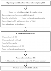
Figure 1

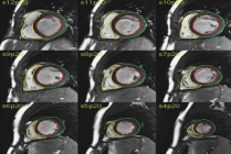
Figure 2

Figure 3
Similar Articles
-
Evaluation of the predictive value of CHA2DS2-VASc Score for no-reflow phenomenon in patients with ST-segment elevation myocardial infarction who underwent Primary Percutaneous Coronary InterventionMahmoud Shawky Abd El-Moneum*. Evaluation of the predictive value of CHA2DS2-VASc Score for no-reflow phenomenon in patients with ST-segment elevation myocardial infarction who underwent Primary Percutaneous Coronary Intervention. . 2019 doi: 10.29328/journal.jccm.1001061; 4: 171-176
-
Evaluation of the effect of coronary artery bypass grafting on the right ventricular function using speckle tracking echocardiographyMahmoud Shawky Abdelmoneum,Neama Ali Elmeligy,Elsayed Abdelkhalek Eldarky,Mohamad Mahmoud Mohamad*. Evaluation of the effect of coronary artery bypass grafting on the right ventricular function using speckle tracking echocardiography. . 2019 doi: 10.29328/journal.jccm.1001075; 4: 236-241
-
Incidence and outcome of no flow after primary percutaneous coronary intervention in acute myocardial infarctionGoutam Datta*. Incidence and outcome of no flow after primary percutaneous coronary intervention in acute myocardial infarction. . 2020 doi: 10.29328/journal.jccm.1001102; 5: 153-156
-
RV Function by cardiac magnetic resonance and its relationship to RV longitudinal strain and neutrophil/lymphocyte ratio in patients with acute inferior ST-segment elevation myocardial infarction undergoing primary percutaneous interventionSalma Taha*,Shrouk Kelany Ali,Fabrizio D’Ascenzo,Hosam Hasan-Ali,Yousra Ghzally#,Mohamed Abdel Ghany. RV Function by cardiac magnetic resonance and its relationship to RV longitudinal strain and neutrophil/lymphocyte ratio in patients with acute inferior ST-segment elevation myocardial infarction undergoing primary percutaneous intervention. . 2021 doi: 10.29328/journal.jccm.1001120; 6: 059-065
-
Isolated multiple pericardial hydatid cysts in an asymptomatic patient: Role of the CMRAntonio Bisignani,Andrea Madeo*,Silvana De Bonis,Riccardo Vico,Giovanni Bisignani. Isolated multiple pericardial hydatid cysts in an asymptomatic patient: Role of the CMR. . 2023 doi: 10.29328/journal.jccm.1001146; 8: 001-003
Recently Viewed
-
Pneumothorax as Complication of CT Guided Lung Biopsy: Frequency, Severity and Assessment of Risk FactorsGaurav Raj*,Neha Kumari,Neha Singh,Kaustubh Gupta,Anurag Gupta,Pradyuman Singh,Hemant Gupta. Pneumothorax as Complication of CT Guided Lung Biopsy: Frequency, Severity and Assessment of Risk Factors. J Radiol Oncol. 2025: doi: 10.29328/journal.jro.1001075; 9: 012-016
-
The Police Power of the National Health Surveillance Agency – ANVISADimas Augusto da Silva*,Rafaela Marinho da Silva. The Police Power of the National Health Surveillance Agency – ANVISA. Arch Cancer Sci Ther. 2024: doi: 10.29328/journal.acst.1001046; 8: 063-076
-
A Comparative Study of Serum Sodium and Potassium Levels across the Three Trimesters of PregnancyOtoikhila OC and Seriki SA*. A Comparative Study of Serum Sodium and Potassium Levels across the Three Trimesters of Pregnancy. Clin J Obstet Gynecol. 2023: doi: 10.29328/journal.cjog.1001137; 6: 108-116
-
Chaos to Cosmos: Quantum Whispers and the Cosmic GenesisOwais Farooq*,Romana Zahoor*. Chaos to Cosmos: Quantum Whispers and the Cosmic Genesis. Int J Phys Res Appl. 2025: doi: 10.29328/journal.ijpra.1001107; 8: 017-023
-
Phytochemical Compounds and the Antifungal Activity of Centaurium pulchellum Ethanol Extracts in IraqNoor Jawad Khadhum, Neepal Imtair Al-Garaawi*, Antethar Jabbar Al-Edani. Phytochemical Compounds and the Antifungal Activity of Centaurium pulchellum Ethanol Extracts in Iraq. J Plant Sci Phytopathol. 2024: doi: 10.29328/journal.jpsp.1001137; 8: 079-083
Most Viewed
-
Evaluation of Biostimulants Based on Recovered Protein Hydrolysates from Animal By-products as Plant Growth EnhancersH Pérez-Aguilar*, M Lacruz-Asaro, F Arán-Ais. Evaluation of Biostimulants Based on Recovered Protein Hydrolysates from Animal By-products as Plant Growth Enhancers. J Plant Sci Phytopathol. 2023 doi: 10.29328/journal.jpsp.1001104; 7: 042-047
-
Sinonasal Myxoma Extending into the Orbit in a 4-Year Old: A Case PresentationJulian A Purrinos*, Ramzi Younis. Sinonasal Myxoma Extending into the Orbit in a 4-Year Old: A Case Presentation. Arch Case Rep. 2024 doi: 10.29328/journal.acr.1001099; 8: 075-077
-
Feasibility study of magnetic sensing for detecting single-neuron action potentialsDenis Tonini,Kai Wu,Renata Saha,Jian-Ping Wang*. Feasibility study of magnetic sensing for detecting single-neuron action potentials. Ann Biomed Sci Eng. 2022 doi: 10.29328/journal.abse.1001018; 6: 019-029
-
Pediatric Dysgerminoma: Unveiling a Rare Ovarian TumorFaten Limaiem*, Khalil Saffar, Ahmed Halouani. Pediatric Dysgerminoma: Unveiling a Rare Ovarian Tumor. Arch Case Rep. 2024 doi: 10.29328/journal.acr.1001087; 8: 010-013
-
Physical activity can change the physiological and psychological circumstances during COVID-19 pandemic: A narrative reviewKhashayar Maroufi*. Physical activity can change the physiological and psychological circumstances during COVID-19 pandemic: A narrative review. J Sports Med Ther. 2021 doi: 10.29328/journal.jsmt.1001051; 6: 001-007

HSPI: We're glad you're here. Please click "create a new Query" if you are a new visitor to our website and need further information from us.
If you are already a member of our network and need to keep track of any developments regarding a question you have already submitted, click "take me to my Query."


