Abstract
Research Article
Evidence of woven bone formation in carotid artery plaques
Mirzaie Masoud*, Zaur Guliyev, Michael Schultz, Peter Schwartz, Johann Philipp Addicks and Sheila Fatehpur
Published: 05 January, 2021 | Volume 6 - Issue 1 | Pages: 001-006
Objective: Plaque morphology plays an important prognostic role in the occurrence of cerebrovascular events. Echolucent and heterogeneous plaques, in particular, carry an increased risk of subsequent stroke. Depending on the quality of the plaque echogenicity based on B-mode ultrasound examination, carotid plaques divide into a soft lipid-rich plaque and a hard plaque with calcification. The aim of this study was to investigate structural changes in the basement membrane of different carotid artery plaque types.
Patients and methods: Biopsies were taken from 10 male patients (average age; 75 + 1 years) and 7 females (68 + 3 years). The study population included patients suffering from a filiform stenosis of the carotid artery, 8 patients with acute cerebrovascular events and 9 with asymptomatic stenosis. Scanning electron and polarised light microscopic investigations were carried out on explanted plaques to determine the morphology of calcified areas in vascular lesions.
Results: By means of scanning electron microscopy, multiple foci of local calcification were identified. The endothelial layer was partially desquamated from the basement membrane and showed island-like formations. Polarised light microscopy allows us to distinguish between soft plaques with transparent structure and hard plaques with woven bone formation.
Conclusion: The major finding of our study is the presence of woven bone tissue in hard plaques of carotid arteries, which may result from pathological strains or mechanical overloading of the collagen fibers. These data suggest a certain parallel with sclerosis of human aortic valves due to their similar morphological characteristics.
Read Full Article HTML DOI: 10.29328/journal.jccm.1001108 Cite this Article Read Full Article PDF
Keywords:
Woven bone; Carotis artery plaques
References
- Moroni F, Ammirati E, Norata GD, Magnoni M, Camici PG. The role of monocytes and macrophages in human atherosclerosis, plaque neoangiogenesis, and atherothrombosis Mediators Inflamm. 2019; 7434376. PubMed: https://pubmed.ncbi.nlm.nih.gov/31089324/
- Zhu G, Hom J, Li Y, Jiang B, Rodriguez F, et al. Carotid plaque imaging and the risk of atherosclerotic cardiovascular disease. Cardiovasc Diagn Ther. 2020; 10: 1048–1067. PubMed: https://pubmed.ncbi.nlm.nih.gov/32968660/
- Johri AM, Nambi V, Naqvi TZ, Feinstein SB, Kim ESH, et al. Recommendations for the assessment of carotid arterial plaque by ultrasound for the characterization of atherosclerosis and evaluation of cardiovascular risk. JASE. 2020; 33: 917-933. PubMed: https://pubmed.ncbi.nlm.nih.gov/32600741/
- Momjian, S, Lalive, P, Sztajzel, R. Carotid plaque: comparison between visual and grey-scale median analysis. Ultrasound Med Biol. 2003; 29: 961-966. PubMed: https://pubmed.ncbi.nlm.nih.gov/12878241/
- Mughal MM, Khan MK, DeMarco JK. Symptomatic and asymptomatic carotid artery plaque, Expert Rev Cardiovasc Ther. 2011; 9: 1315–1330. PubMed: https://www.ncbi.nlm.nih.gov/pmc/articles/PMC3243497/
- Mirzaie M, Schultz M, Schwartz P. Evidence of woven bone formation in heart valve disease. Ann Thorac Cardiovasc Surg. 2003; 9: 163-169. PubMed: https://pubmed.ncbi.nlm.nih.gov/12875637/
- Von Hagens G. Impregnation of soft biological specimens with thermostetting resins and elastomers. Anat Rec. 1979; 94: 247–255. PubMed: https://pubmed.ncbi.nlm.nih.gov/464325/
- Schultz M, Drommer R. Possibilities of preparation production from the craniofacial area for macroscopic and microscopic examination using new plastics techniques. Exp Mund-KieferGesichts Chir. 1983; 28: 95–97.
- Meershoek A, van Dijk RA, Verhage S. Hamming JF, van den Bogaerdt AJ, et al. Histological evaluation disqualifies IMT and calcification scores as surrogates for grading coronary and aortic atherosclerosis. Int J Cardiol. 2016; 224: 328-334. PubMed: https://pubmed.ncbi.nlm.nih.gov/27668706/
- Nambi V, Chambless L, Folsom AR. He M, Hu Y, et al. Carotid intima-media thickness and presence or absence of plaque improves prediction of coronary heart disease risk: The ARIC (Atherosclerosis risk in communities) study. J Am Coll Cardiol. 2010; 55: 1600-1607. PubMed: https://pubmed.ncbi.nlm.nih.gov/20378078/
- Toutouzas, K, Karanasos, A, Tousoulis, D. Optical coherence tomography for the detection of the vulnerable plaque. Eur Cardiol. 2016; 11: 90–95. PubMed: https://pubmed.ncbi.nlm.nih.gov/30310454/
- Hua Y. Chinese guidance on vascular ultrasound examination in stroke. Chin J Med Ultrasound. 2015; 8: 599-610.
- Pelisek J, Wendorff H, Wendorff C. Age-associated changes in human carotid atherosclerotic plaques. Ann Med. 2016; 48: 541-551. PubMed: https://pubmed.ncbi.nlm.nih.gov/27595161/
- Jinnouchi H, Sato Y, Sakamoto A, Cornelissen A, Mori M, et al. Calcium deposition within coronary atherosclerotic lesion: Implications for plaque stability. Finn Atherosclerosis. 2020; 306: 85-95. PubMed: https://pubmed.ncbi.nlm.nih.gov/32654790/
- Antipova D, Eadie L, Makin S. The use of transcranial ultrasound and clinical assessment to diagnose ischaemic stroke due to large vessel occlusion in remote and rural areas, PLoS One. 2020; 15: e0239653. PubMed: https://pubmed.ncbi.nlm.nih.gov/33007053/
- Ahluwalia N, Genoux A, Ferrieres J, Perret B, Carayol M, et al. Iron status is associated with carotid atherosclerotic plaques in middle-aged adults. J Nutr. 2010; 140: 812–816. PubMed: https://pubmed.ncbi.nlm.nih.gov/20181783/
- Sun B, Zhao H, Liu X, Lu Q, Zhao X, et al. Elevated hemoglobin A1c is associated with carotid plaque vulnerability: Novel findings from magnetic resonance imaging study in hypertensive stroke patients. Sci Rep. 2016; 6: 33246. PubMed: https://pubmed.ncbi.nlm.nih.gov/27629481/
- Brophy ML, Dong Y, Wu H, Rahman HNA, Song K, et al. Eating the dead to keep atherosclerosis at bay. Front Cardiovasc Med. 2017; 4: 2. PubMed: https://pubmed.ncbi.nlm.nih.gov/28194400/
- Vaidya K, Martínez G, Patel S. The role of colchicine in acute coronary syndromes. Clin Ther. 2019; 41: 11-20. PubMed: https://pubmed.ncbi.nlm.nih.gov/30185392/
- Ruddy JM, Ikonomidis JS, Jones JA. Multidimensional contribution of matrix metalloproteinases to atherosclerotic plaque vulnerability: Multiple mechanisms of inhibition to promote stability. J Vasc Res. 2016; 53: 1-16. PubMed: https://pubmed.ncbi.nlm.nih.gov/27327039/
- Alhendi AMN, Patrikakis M, Daub CO, Kawaji H, Itoh M, et al. Promoter usage and dynamics in vascular smooth muscle cells Exposed to fibroblast growth factor-2 or interleukin-1β. Sci Rep. 2018; 8: 13164. PubMed: https://pubmed.ncbi.nlm.nih.gov/30177712/
- Veseli BE, Perrotta P, De Meyer GRA, Roth L, der Donckt CV, et al. Animal models of atherosclerosis. Eur J Pharmacol. 2017; 816: 3-13. PubMed: https://pubmed.ncbi.nlm.nih.gov/28483459/
- Nikkari ST, O'Brien KD, Ferguson M, Hatsukami T, Welgus HG, et al. Interstitial collagenase (MMP-1) expression in human carotid atherosclerosis. Circulation. 1995; 92: 1393-1398. PubMed: https://pubmed.ncbi.nlm.nih.gov/7664418/
- Chen X, Cao X, Jiang H, Che X, Xu X, et al. SIKVAV-modified chitosan hydrogel as a skin substitutes for wound closure in mice. Molecules. 2018; 23: 2611. PubMed: https://pubmed.ncbi.nlm.nih.gov/30314388/
- Cheng S, Wang D, Jin Ke J, Ma L, Zhou J, et al. Improved in vitro angiogenic behavior of human umbilical vein endothelial cells with oxidized polydopamine coating. Colloid Surface B. 2020; 194: 111176. PubMed: https://pubmed.ncbi.nlm.nih.gov/32540767/
- Chia JS, Du JL, Hsu WB, Sun A, Chiang CP, et al. Inhibition of metastasis, angiogenesis, and tumor growth by Chinese herbal cocktail Tien-Hsien Liquid. BMC Cancer. 2010; 10: 175. PubMed: https://pubmed.ncbi.nlm.nih.gov/20429953/
- Latha S, Samiappan D, Kumar R. Carotid artery ultrasound image analysis: A review of the literature. Proc Inst Mech Eng H. 2020; 234: 417-443. PubMed: https://pubmed.ncbi.nlm.nih.gov/31960771/
- Saxena A, Kwee Ng EY, Teik S. Imaging modalities to diagnose carotid artery stenosis: progress and prospect, Bio Med Eng. 2019; 18: 66. PubMed: https://pubmed.ncbi.nlm.nih.gov/31138235/
- Banaei A. The comparison between digital subtraction angiography, CT angiography, and doppler ultrasonography in evaluation and assessment of carotid artery stenosis. Ann Mil Health Sci Res. 2017; 15: e61661.
- Correia M, Maresca D, Goudot G, Villemain O, Bizé A, et al. Quantitative imaging of coronary flows using 3D ultrafast doppler coronary angiography. Phys Med Biol. 2020; 65: 105013. PubMed: https://pubmed.ncbi.nlm.nih.gov/32340010/
- Morbiducci U, Kok AM, Kwak BR. Atherosclerosis at arterial bifurcations: Evidence for the role of haemodynamics and geometry. Thrombosis and Haemostasis. 2016; 115: 484492. PubMed: https://pubmed.ncbi.nlm.nih.gov/26740210/
- Monckeberg JG. Normal histological construction and sclerosis of aortic valves. Virchows Archiv Pathol Anat Physiol. 1904; 176: 472-514.
- Virchow R. Cellular pathology: as based upon physiological and pathological histology. Nutr Rev. 1989; 47: 23-25. PubMed: https://pubmed.ncbi.nlm.nih.gov/2649802/
- Yang P, Troncone L, Augur ZM, Kim SSJ, McNeil ME, et al. The role of bone morphogenetic protein signaling in vascular calcification. Bone. 2020; 115542. PubMed: https://pubmed.ncbi.nlm.nih.gov/32736145/
- Sun M, Chang Q, Xin M. Endogenous bone morphogenetic protein 2 plays a role in vascular smooth muscle cell calcification induced by interleukin 6 in vitro. Int J Immunopathol Pharmacol. 2017; 30: 227–237. PubMed: https://pubmed.ncbi.nlm.nih.gov/28134597/
- Nagy E, Eriksson P, Yousry M, Caidahl K, Ingelsson E, et al. Valvular osteoclasts in calcification and aortic valve stenosis severity. Int J Cardiol. 2013; 168: 2264-2271. PubMed: https://pubmed.ncbi.nlm.nih.gov/23452891/
- Low EL, Baker AH, Bradshaw AC. TGFβ, smooth muscle cells and coronary artery disease: a review. Cell Signal. 2019; 53: 90-101. PubMed: https://pubmed.ncbi.nlm.nih.gov/30227237/
- Shioi A, Ikari Y. Plaque calcification during atherosclerosis progression and regression. J Atheroscler Thromb. 2018; 25: 294–303. PubMed: https://pubmed.ncbi.nlm.nih.gov/29238011/
Figures:
Figure 1
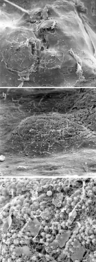
Figure 2
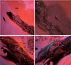
Figure 3
Similar Articles
-
Evidence of woven bone formation in carotid artery plaquesMirzaie Masoud*,Zaur Guliyev,Michael Schultz,Peter Schwartz,Johann Philipp Addicks,Sheila Fatehpur. Evidence of woven bone formation in carotid artery plaques. . 2021 doi: 10.29328/journal.jccm.1001108; 6: 001-006
Recently Viewed
-
The lateralization pattern has an influence on the severity of ankle sprainsAndrzej Mioduszewski*, Mikołaj Wróbel, Emilia Hammar. The lateralization pattern has an influence on the severity of ankle sprains. J Sports Med Ther. 2023: doi: 10.29328/journal.jsmt.1001066; 8: 016-020
-
Role of RBC Parameters to Differentiate between Iron Deficiency Anemia and Anemia of Chronic DiseasesReena Bhaisare*, Ravindranath M, Gurmeet Singh. Role of RBC Parameters to Differentiate between Iron Deficiency Anemia and Anemia of Chronic Diseases. J Hematol Clin Res. 2023: doi: 10.29328/journal.jhcr.1001024; 7: 021-024
-
Non-invasive Serological Markers of Hepatic Fibrosis – Mini ReviewElena Popa*, Raluca Ioana Avram, Andrei Emilian Popa, Adorata Elena Coman. Non-invasive Serological Markers of Hepatic Fibrosis – Mini Review. Arch Surg Clin Res. 2024: doi: 10.29328/journal.ascr.1001081; 8: 032-038
-
Knowledge of diabetic patients regarding diabetes management, diet, lifestyle modification and blood glucose monitoringZarmeena Qazi*,Hafiz Muhammad Dawood. Knowledge of diabetic patients regarding diabetes management, diet, lifestyle modification and blood glucose monitoring. Arch Pharm Pharma Sci. 2023: doi: 10.29328/journal.apps.1001036; 7: 001-003
-
How does a Personalized Rehabilitative Model influence the Functional Response of Different Ankle Foot Orthoses in a Cohort of Patients Affected by Neurological Gait Pattern?Maurizio Falso*, Eleonora Cattaneo,Elisa Foglia,Marco Zucchini,Franco Zucchini. How does a Personalized Rehabilitative Model influence the Functional Response of Different Ankle Foot Orthoses in a Cohort of Patients Affected by Neurological Gait Pattern?. J Nov Physiother Rehabil. 2017: doi: 10.29328/journal.jnpr.1001010; 1: 072-092
Most Viewed
-
Evaluation of Biostimulants Based on Recovered Protein Hydrolysates from Animal By-products as Plant Growth EnhancersH Pérez-Aguilar*, M Lacruz-Asaro, F Arán-Ais. Evaluation of Biostimulants Based on Recovered Protein Hydrolysates from Animal By-products as Plant Growth Enhancers. J Plant Sci Phytopathol. 2023 doi: 10.29328/journal.jpsp.1001104; 7: 042-047
-
Sinonasal Myxoma Extending into the Orbit in a 4-Year Old: A Case PresentationJulian A Purrinos*, Ramzi Younis. Sinonasal Myxoma Extending into the Orbit in a 4-Year Old: A Case Presentation. Arch Case Rep. 2024 doi: 10.29328/journal.acr.1001099; 8: 075-077
-
Feasibility study of magnetic sensing for detecting single-neuron action potentialsDenis Tonini,Kai Wu,Renata Saha,Jian-Ping Wang*. Feasibility study of magnetic sensing for detecting single-neuron action potentials. Ann Biomed Sci Eng. 2022 doi: 10.29328/journal.abse.1001018; 6: 019-029
-
Pediatric Dysgerminoma: Unveiling a Rare Ovarian TumorFaten Limaiem*, Khalil Saffar, Ahmed Halouani. Pediatric Dysgerminoma: Unveiling a Rare Ovarian Tumor. Arch Case Rep. 2024 doi: 10.29328/journal.acr.1001087; 8: 010-013
-
Physical activity can change the physiological and psychological circumstances during COVID-19 pandemic: A narrative reviewKhashayar Maroufi*. Physical activity can change the physiological and psychological circumstances during COVID-19 pandemic: A narrative review. J Sports Med Ther. 2021 doi: 10.29328/journal.jsmt.1001051; 6: 001-007

HSPI: We're glad you're here. Please click "create a new Query" if you are a new visitor to our website and need further information from us.
If you are already a member of our network and need to keep track of any developments regarding a question you have already submitted, click "take me to my Query."


