Abstract
Review Article
Glycosaminoglycans as Novel Targets for in vivo Contrast-Enhanced Magnetic Resonance Imaging of Atherosclerosis
Yavuz O Uca* and Matthias Taupitz
Published: 20 April, 2020 | Volume 5 - Issue 1 | Pages: 080-088
Atherosclerosis is an important promoter of cardiovascular disease potentiating myocardial infarction or stroke. Current demand in biomedical imaging necessitates noninvasive characterization of arterial changes responsible for transition of stable plaque into rupture-prone vulnerable plaque. in vivo contrast enhanced magnetic resonance (MR) imaging (MRI) allows quantitative and functional monitoring of pathomorphological changes through signal differences induced by the contrast agent uptake in the diseased vessel wall, therefore it is the ideal modality toward this goal. However, studies have so far focused on the cellular targets of persisting inflammation, leaving extracellular matrix (ECM) far behind. In this review, we portray ECM remodeling during atherosclerotic plaque progression by summarizing the state of the-art in MRI and current imaging targets. Finally, we aim to discuss glycosaminoglycans (GAGs) and their functional interactions, which might offer potential toward development of novel imaging probes for in vivo contrast-enhanced MRI of atherosclerosis.
Read Full Article HTML DOI: 10.29328/journal.jccm.1001091 Cite this Article Read Full Article PDF
References
- Ross R. Atherosclerosis — An Inflammatory Disease. N Engl J Med. 1999; 340: 115–126. PubMed: https://www.ncbi.nlm.nih.gov/pubmed/9887164
- Benjamin EJ, Muntner P, Alonso A, Bittencourt MS, Callaway CW, et al. Heart disease and stroke statistics-2019 update: a report From the American Heart Association. Circulation. 2019; 139 :526–528. PubMed: https://www.ncbi.nlm.nih.gov/pubmed/30700139
- Nichols M, Townsend N, Scarborough P, Rayner M. Cardiovascular disease in Europe 2014: epidemiological update. Eur Heart J. 2014; 35: 2950–2959. PubMed: https://www.ncbi.nlm.nih.gov/pubmed/25139896
- Heidenreich PA, Trogdon JG, Khavjou OA, Butler J, Dracup K, et al. Forecasting the Future of Cardiovascular Disease in the United States: A Policy Statement From the American Heart Association. Circulation. 2011; 123: 933–944. PubMed: https://www.ncbi.nlm.nih.gov/pubmed/21262990
- Nojiri S, Daida H. Atherosclerotic Cardiovascular Risk in Japan. Jpn Clin Med [Internet]. 2017; 8. PubMed: http://www.ncbi.nlm.nih.gov/pmc/articles/PMC5480958/
- Pozo E, Agudo-Quilez P, Rojas-González A, Alvarado T, Olivera MJ, et al. Noninvasive diagnosis of vulnerable coronary plaque. World J Cardiol. 2016; 8: 520–533. PubMed: https://www.ncbi.nlm.nih.gov/pmc/articles/PMC5039354/
- Tarkin JM, Dweck MR, Evans NR, Takx RA, Brown AJ, et al. Imaging atherosclerosis. Circ Res. 2016; 118: 750–769. PubMed: https://www.ncbi.nlm.nih.gov/pubmed/26892971
- Skinner MP, Yuan C, Mitsumori L, Hayes CE, M.p.s EWR, Nelson JA, et al. Serial magnetic resonance imaging of experimental atherosclerosis detects lesion fine structure, progression and complications in vivo. Nat Med. 1995; 1: 69–73. PubMed: https://www.ncbi.nlm.nih.gov/pubmed/7584956
- Toussaint J-F, LaMuraglia GM, Southern JF, Fuster V, Kantor HL. Magnetic Resonance Images Lipid, Fibrous, Calcified, Hemorrhagic, and Thrombotic Components of Human Atherosclerosis in vivo. Circulation. 1996; 94: 932–8.
- Botnar RM, Stuber M, Kissinger KV, Kim WY, Spuentrup E, et al. Noninvasive Coronary Vessel Wall and Plaque Imaging With Magnetic Resonance Imaging. Circulation. 2000; 102: 2582–2587. PubMed: https://www.ncbi.nlm.nih.gov/pubmed/11085960
- Fayad ZA, Fuster V. Clinical Imaging of the High-Risk or Vulnerable Atherosclerotic Plaque. Circ Res. 2001; 89: 305–316. PubMed: https://www.ncbi.nlm.nih.gov/pubmed/11509446
- Tang TY, Muller KH, Graves MJ, Li ZY, Walsh SR, et al. Iron Oxide Particles for Atheroma Imaging. Arterioscler Thromb Vasc Biol. 2009; 29:1001–1008. PubMed: https://www.ncbi.nlm.nih.gov/pubmed/19229073
- Falk E, Shah PK, Fuster V. Coronary Plaque Disruption. Circulation. 1995; 92: 657–671. PubMed: https://www.ncbi.nlm.nih.gov/pubmed/7634481
- Virmani R, Kolodgie FD, Burke AP, Finn AV, Gold HK, N T, et al. Atherosclerotic Plaque Progression and Vulnerability to Rupture.
- Hansson GK. Inflammation, Atherosclerosis, and Coronary Artery Disease. N Engl J Med. 2005; 352: 1685–1695. PubMed: https://www.ncbi.nlm.nih.gov/pubmed/15843671
- Libby P, DiCarli M, Weissleder R. The Vascular Biology of Atherosclerosis and Imaging Targets. J Nucl Med. 2010; 51(Supplement 1): 335-375. PubMed: https://www.ncbi.nlm.nih.gov/pubmed/20395349
- Rudd JHF, Hyafil F, Fayad ZA. Inflammation Imaging in Atherosclerosis. Arterioscler Thromb Vasc Biol. 2009; 29: 1009–1016.
- Reimann C, Brangsch J, Colletini F, Walter T, Hamm B, Botnar RM, et al. Molecular imaging of the extracellular matrix in the context of atherosclerosis. Adv Drug Deliv Rev. 2017; 113:49–60. PubMed: https://www.ncbi.nlm.nih.gov/pubmed/27639968
- Virmani R, Burke AP, Farb A, Kolodgie FD. Pathology of the Vulnerable Plaque. J Am Coll Cardiol. 2006; 47(8 Supplement): C13–18. PubMed: https://www.ncbi.nlm.nih.gov/pubmed/16631505
- Järveläinen H, Sainio A, Koulu M, Wight TN, Penttinen R. Extracellular Matrix Molecules: Potential Targets in Pharmacotherapy. Pharmacol Rev. 2009; 61:198–223. PubMed: https://www.ncbi.nlm.nih.gov/pmc/articles/PMC2830117/s
- Lutolf MP, Hubbell JA. Synthetic biomaterials as instructive extracellular microenvironments for morphogenesis in tissue engineering. Nat Biotechnol. 2005; 23: 47–55. PubMed: https://www.ncbi.nlm.nih.gov/pubmed/15637621
- Grobner T. Gadolinium – a specific trigger for the development of nephrogenic fibrosing dermopathy and nephrogenic systemic fibrosis? Nephrol Dial Transplant. 2006; 21: 1104–1108. PubMed: https://www.ncbi.nlm.nih.gov/pubmed/16431890
- George S j., Webb S m., Abraham J l., Cramer S p. Synchrotron X-ray analyses demonstrate phosphate-bound gadolinium in skin in nephrogenic systemic fibrosis. Br J Dermatol. 2010; 163: 1077–1081. PubMed: https://www.ncbi.nlm.nih.gov/pubmed/20560953
- Laurent S, Vander Elst L, Henoumont C, Muller RN. How to measure the transmetallation of a gadolinium complex. Contrast Media Mol Imaging. 2010; 5: 305–308. PubMed: https://www.ncbi.nlm.nih.gov/pubmed/20803503
- Taupitz M, Stolzenburg N, Ebert M, Schnorr J, Hauptmann R, Kratz H, et al. Gadolinium-containing magnetic resonance contrast media: investigation on the possible transchelation of Gd 3+ to the glycosaminoglycan heparin: GdCM, Glycosaminoglycans and Transchelation. Contrast Media Mol Imaging. 2013; 8:108–116. PubMed: https://www.ncbi.nlm.nih.gov/pubmed/23281283
- Narula J, Virmani R, Iskandrian AE. Strategic targeting of atherosclerotic lesions. J Nucl Cardiol. 1999; 6: 81–90. PubMed: https://www.ncbi.nlm.nih.gov/pubmed/10070844
- Chatzizisis YS, Coskun AU, Jonas M, Edelman ER, Feldman CL, et al. Role of Endothelial Shear Stress in the Natural History of Coronary Atherosclerosis and Vascular Remodeling: Molecular, Cellular, and Vascular Behavior. J Am Coll Cardiol. 2007; 49: 2379–2393. PubMed: https://www.ncbi.nlm.nih.gov/pubmed/17599600
- Gimbrone MA, Topper JN, Nagel T, Anderson KR, Garcia-Cardeña G. Endothelial Dysfunction, Hemodynamic Forces, and Atherogenesisa. Ann N Y Acad Sci. 2000; 902: 230–240. PubMed: https://www.ncbi.nlm.nih.gov/pubmed/10865843
- Navab M, Ananthramaiah GM, Reddy ST, Van Lenten BJ, Ansell BJ, et al. The Pathogenesis of Atherosclerosis-The oxidation hypothesis of atherogenesis: the role of oxidized phospholipids and HDL. J Lipid Res. 2004; 45: 993–1007. PubMed: https://www.ncbi.nlm.nih.gov/pubmed/15060092
- Blankenberg S, Barbaux S, Tiret L. Adhesion molecules and atherosclerosis. Atherosclerosis. 2003; 170: 191–203. PubMed: https://www.ncbi.nlm.nih.gov/pubmed/14612198
- Winter PM, Morawski AM, Caruthers SD, Fuhrhop RW, Zhang H, et al. Molecular Imaging of Angiogenesis in Early-Stage Atherosclerosis With αvβ3-Integrin–Targeted Nanoparticles. Circulation. 2003; 108: 2270–2274. PubMed: https://www.ncbi.nlm.nih.gov/pubmed/14557370
- Galkina E, Ley K. Vascular Adhesion Molecules in Atherosclerosis. Arterioscler Thromb Vasc Biol. 2007; 27: 2292–2301. PubMed: https://www.ncbi.nlm.nih.gov/pubmed/17673705
- Kelly KA, Allport JR, Tsourkas A, Shinde-Patil VR, Josephson L, et al. Detection of Vascular Adhesion Molecule-1 Expression Using a Novel Multimodal Nanoparticle. Circ Res. 2005; 96: 327–336. PubMed: https://www.ncbi.nlm.nih.gov/pubmed/15653572
- Nahrendorf M, Jaffer FA, Kelly KA, Sosnovik DE, Aikawa E, et al. Noninvasive Vascular Cell Adhesion Molecule-1 Imaging Identifies Inflammatory Activation of Cells in Atherosclerosis. Circulation. 2006; 114: 1504–1511. PubMed: https://www.ncbi.nlm.nih.gov/pubmed/17000904
- Briley-Saebo KC, Shaw PX, Mulder WJM, Choi S-H, Vucic E, et al. Targeted Molecular Probes for Imaging Atherosclerotic Lesions With Magnetic Resonance Using Antibodies That Recognize Oxidation-Specific Epitopes. Circulation. 2008; 117: 3206–3215. PubMed: https://www.ncbi.nlm.nih.gov/pubmed/18541740
- McAteer MA, Schneider JE, Ali ZA, Warrick 1 Nicholas, Bursill CA, et al. Magnetic Resonance Imaging of Endothelial Adhesion Molecules in Mouse Atherosclerosis Using Dual-Targeted Microparticles of Iron Oxide. Arterioscler Thromb Vasc Biol. 2008 Jan;28(1):77–83. PubMed: https://www.ncbi.nlm.nih.gov/pubmed/17962629
- Kwon HM, Sangiorgi G, Ritman EL, McKenna C, Holmes DR, et al. Enhanced coronary vasa vasorum neovascularization in experimental hypercholesterolemia. J Clin Invest. 1998; 101: 1551–1556. PubMed: https://www.ncbi.nlm.nih.gov/pubmed/9541483
- Libby P. Inflammation in atherosclerosis. Nature. 2002; 420: 868–874.
- Amirbekian V, Lipinski MJ, Briley-Saebo KC, Amirbekian S, Aguinaldo JGS, et al. Detecting and assessing macrophages in vivo to evaluate atherosclerosis noninvasively using molecular MRI. Proc Natl Acad Sci. 2007; 104: 961–966. PubMed: https://www.ncbi.nlm.nih.gov/pubmed/17215360
- Ruehm SG, Corot C, Vogt P, Kolb S, Debatin JF. Magnetic Resonance Imaging of Atherosclerotic Plaque With Ultrasmall Superparamagnetic Particles of Iron Oxide in Hyperlipidemic Rabbits. Circulation. 2001; 103: 415–422. PubMed: https://www.ncbi.nlm.nih.gov/pubmed/11157694
- Schmitz SA, Taupitz M, Wagner S, Wolf K-J, Beyersdorff D, et al. Magnetic resonance imaging of atherosclerotic plaques using superparamagnetic iron oxide particles. J Magn Reson Imaging. 2001; 14: 355–361. PubMed: https://www.ncbi.nlm.nih.gov/pubmed/11599058
- Kooi ME, Cappendijk VC, Cleutjens KBJM, Kessels AGH, Kitslaar PJEHM, et al. Accumulation of Ultrasmall Superparamagnetic Particles of Iron Oxide in Human Atherosclerotic Plaques Can Be Detected by in vivo Magnetic Resonance Imaging. Circulation. 2003; 107: 2453–2458. PubMed: https://www.ncbi.nlm.nih.gov/pubmed/12719280
- Chen J, Tung CH, Mahmood U, Ntziachristos V, Gyurko R, et al. in vivo Imaging of Proteolytic Activity in Atherosclerosis. Circulation. 2002; 105: 2766–2771. PubMed: https://www.ncbi.nlm.nih.gov/pubmed/12057992
- Deguchi J, Aikawa M, Tung C-H, Aikawa E, Kim D-E, et al. Inflammation in Atherosclerosis: Visualizing Matrix Metalloproteinase Action in Macrophages in vivo. Circulation. 2006 Jul 4;114(1):55–62. PubMed: https://www.ncbi.nlm.nih.gov/pubmed/16801460
- Shipley JM, Wesselschmidt RL, Kobayashi DK, Ley TJ, Shapiro SD. Metalloelastase is required for macrophage-mediated proteolysis and matrix invasion in mice. Proc Natl Acad Sci. 1996; 93: 3942–3946. PubMed: https://www.ncbi.nlm.nih.gov/pmc/articles/PMC39464/
- Hyafil F, Vucic E, Cornily J-C, Sharma R, Amirbekian V, et al. Monitoring of arterial wall remodelling in atherosclerotic rabbits with a magnetic resonance imaging contrast agent binding to matrix metalloproteinases. Eur Heart J. 2011; 32: 1561–1571. PubMed: https://www.ncbi.nlm.nih.gov/pubmed/21118852
- Helske S, Syväranta S, Lindstedt KA, Lappalainen J, Öörni K, et al. Increased Expression of Elastolytic Cathepsins S, K, and V and Their Inhibitor Cystatin C in Stenotic Aortic Valves. Arterioscler Thromb Vasc Biol. 2006; 26: 1791–1798. PubMed: https://www.ncbi.nlm.nih.gov/pubmed/16728655
- Majmudar MD, Nahrendorf M. Cardiovascular Molecular Imaging: The Road Ahead. J Nucl Med. 2012; 53: 673–676. PubMed: https://www.ncbi.nlm.nih.gov/pubmed/22492729
- Lairez O, Fayad ZA. Imaging of atherosclerosis: Can molecular imaging do more? Arch Cardiovasc Dis. 2013; 106: 551–553.
- Carlier S, Kakadiaris IA, Dib N, Vavuranakis M, O’Malley SM, et al. Vasa vasorum imaging: A new window to the clinical detection of vulnerable atherosclerotic plaques. Curr Atheroscler Rep. 2005; 7: 164–169. PubMed: https://www.ncbi.nlm.nih.gov/pubmed/15727733
- Vakonakis I, Campbell ID. Extracellular matrix: from atomic resolution to ultrastructure. Curr Opin Cell Biol. 2007; 19: 578–583. PubMed: https://www.ncbi.nlm.nih.gov/pubmed/17942296
- Aumailley M, Gayraud B. Structure and biological activity of the extracellular matrix. J Mol Med. 1998; 76: 253–265. PubMed: https://www.ncbi.nlm.nih.gov/pubmed/9535559
- Katsuda S, Kaji T. Atherosclerosis and Extracellular Matrix. J Atheroscler Thromb. 2003; 10: 267–274. PubMed: https://www.ncbi.nlm.nih.gov/pubmed/14718743
- Wight TN, Lara S, Riessen R, Le Baron R, Isner J. Selective deposits of versican in the extracellular matrix of restenotic lesions from human peripheral arteries. Am J Pathol. 1997; 151: 963-973. PubMed: https://www.ncbi.nlm.nih.gov/pubmed/9327730
- Wight TN, Merrilees MJ. Proteoglycans in Atherosclerosis and Restenosis: Key Roles for Versican. Circ Res. 2004; 94: 1158–1167. PubMed: https://www.ncbi.nlm.nih.gov/pubmed/15142969
- Matter CM, Schuler PK, Alessi P, Meier P, Ricci R, Zhang D, et al. Molecular Imaging of Atherosclerotic Plaques Using a Human Antibody Against the Extra-Domain B of Fibronectin. Circ Res. 2004; 95: 1225–1233.
- Ye F, Jeong E-K, Jia Z, Yang T, Parker D, et al. A Peptide Targeted Contrast Agent Specific to Fibrin-Fibronectin Complexes for Cancer Molecular Imaging with MRI. Bioconjug Chem. 2008; 19: 2300–2303. PubMed: https://www.ncbi.nlm.nih.gov/pubmed/19053180
- Mayne R. Collagenous proteins of blood vessels. Arterioscler Thromb Vasc Biol. 1986; 6: 585–593.
- Brodsky B, Persikov AV. Molecular Structure of the Collagen Triple Helix. Adv Protein Chem. 2005; 70: 301–339. PubMed: https://www.ncbi.nlm.nih.gov/pubmed/15837519
- Caravan P, Das B, Dumas S, Epstein FH, Helm PA, et al. Collagen-Targeted MRI Contrast Agent for Molecular Imaging of Fibrosis. Angew Chem. 2007; 119: 8319–8321. PubMed: https://www.ncbi.nlm.nih.gov/pubmed/17893943
- Helm PA, Caravan P, French BA, Jacques V, Shen L, Xu Y, et al. Postinfarction myocardial scarring in mice: molecular MR imaging with use of a collagen-targeting contrast agent. Radiology. 2008; 247: 788–796. PubMed: https://www.ncbi.nlm.nih.gov/pmc/articles/PMC5410958/
- Jimi S, Sakata N, Matunaga A, Takebayashi S. Low density lipoproteins bind more to type I and III collagens by negative charge-dependent mechanisms than to type IV and V collagens. Atherosclerosis. 1994; 107: 109–116. PubMed: https://www.ncbi.nlm.nih.gov/pubmed/7945553
- Khalil MF. Molecular Interactions Leading to Lipoprotein Retention and the Initiation of Atherosclerosis. Arterioscler Thromb Vasc Biol. 2004; 24: 2211–2218. PubMed: https://www.ncbi.nlm.nih.gov/pubmed/15472124
- Adiguzel E, Ahmad PJ, Franco C, Bendeck MP. Collagens in the progression and complications of atherosclerosis. Vasc Med. 2009; 14: 73–89. PubMed: https://www.ncbi.nlm.nih.gov/pubmed/19144782
- Chen W, Cormode DP, Vengrenyuk Y, Herranz B, Feig JE, et al. Collagen-Specific Peptide Conjugated HDL Nanoparticles as MRI Contrast Agent to Evaluate Compositional Changes in Atherosclerotic Plaque Regression. JACC Cardiovasc Imaging. 2013; 6: 373–384. PubMed: https://www.ncbi.nlm.nih.gov/pubmed/23433925
- Spuentrup E, Ruhl KM, Botnar RM, Wiethoff AJ, Buhl A, et al. Molecular Magnetic Resonance Imaging of Myocardial Perfusion With EP-3600, a Collagen-Specific Contrast Agent: Initial Feasibility Study in a Swine Model. Circulation. 2009; 119: 1768–1775. PubMed: https://www.ncbi.nlm.nih.gov/pubmed/19307474
- Li DY, Brooke B, Davis EC, Mecham RP, Sorensen LK, et al. Elastin is an essential determinant of arterial morphogenesis. Nature. 1998; 393: 276–280. PubMed: https://www.ncbi.nlm.nih.gov/pubmed/9607766
- Karnik SK, Brooke BS, Bayes-Genis A, Sorensen L, Wythe JD, et al. A critical role for elastin signaling in vascular morphogenesis and disease. Development. 2003; 130: 411–423. PubMed: https://www.ncbi.nlm.nih.gov/pubmed/12466207
- Kwon GP, Schroeder JL, Amar MJ, Remaley AT, Balaban RS. Contribution of Macromolecular Structure to the Retention of Low-Density Lipoprotein at Arterial Branch Points. Circulation. 2008; 117: 2919–2927. PubMed: https://www.ncbi.nlm.nih.gov/pubmed/18506002
- Robert L, Robert AM, Jacotot B. Elastin–elastase–atherosclerosis revisited. Atherosclerosis. 1998; 140: 281–295. PubMed: https://www.ncbi.nlm.nih.gov/pubmed/9862271
- Krettek A, Sukhova GK, Libby P. Elastogenesis in Human Arterial Disease: A Role for Macrophages in Disordered Elastin Synthesis. Arterioscler Thromb Vasc Biol. 2003; 23: 582–587. PubMed: https://www.ncbi.nlm.nih.gov/pubmed/12615674
- Choudhury Robin P, Valentin F, Badimon Juan J, Fisher Edward A, Fayad Zahi A. MRI and Characterization of Atherosclerotic Plaque. Arterioscler Thromb Vasc Biol. 2002; 22: 1065–1074. PubMed: https://www.ncbi.nlm.nih.gov/pubmed/12117718
- Makowski MR, Wiethoff AJ, Blume U, Cuello F, Warley A, et al. Assessment of atherosclerotic plaque burden with an elastin-specific magnetic resonance contrast agent. Nat Med. 2011; 17: 383–388. PubMed: https://www.ncbi.nlm.nih.gov/pubmed/21336283
- Fuster V, Stein B, Ambrose JA, Badimon L, Badimon JJ, et al. Atherosclerotic plaque rupture and thrombosis. Evolving concepts. Circulation. 1990; 82(3 Suppl): II47-1159. PubMed: https://www.ncbi.nlm.nih.gov/pubmed/2203564
- La Corte ALC, Philippou H, Ariëns RAS. Role of Fibrin Structure in Thrombosis and Vascular Disease. Adv Protein Chem Struct Biol. 2011; 83: 75–127. PubMed: https://www.ncbi.nlm.nih.gov/pubmed/21570666
- Tavora F, Cresswell N, Li L, Ripple M, Burke A. Immunolocalisation of fibrin in coronary atherosclerosis: implications for necrotic core development. Pathology (Phila). 2010; 42: 15‑ PubMed: https://www.ncbi.nlm.nih.gov/pubmed/20025475
- Botnar RM, Perez AS, Witte S, Wiethoff AJ, Laredo J, et al. in vivo Molecular Imaging of Acute and Subacute Thrombosis Using a Fibrin-Binding Magnetic Resonance Imaging Contrast Agent. Circulation. 2004; 109: 2023–2029. PubMed: https://www.ncbi.nlm.nih.gov/pubmed/15066940
- Makowski MR, Forbes SC, Blume U, Warley A, Jansen CHP, et al. in vivo assessment of intraplaque and endothelial fibrin in ApoE−/− mice by molecular MRI. Atherosclerosis. 2012; 222: 43–49.
- Flacke S, Fischer S, Scott MJ, Fuhrhop RJ, Allen JS, et al. Novel MRI Contrast Agent for Molecular Imaging of Fibrin: Implications for Detecting Vulnerable Plaques. Circulation. 2001; 104: 1280–1285. PubMed: https://www.ncbi.nlm.nih.gov/pubmed/11551880
- Sirol M, Aguinaldo JGS, Graham PB, Weisskoff R, Lauffer R, et al. Fibrin-targeted contrast agent for improvement of in vivo acute thrombus detection with magnetic resonance imaging. Atherosclerosis. 2005; 182: 79–85. PubMed: https://www.ncbi.nlm.nih.gov/pubmed/16115477
- Jackson RL, Busch SJ, Cardin AD. Glycosaminoglucans : molecular properties, protein interactions, and role in physiological processes. Physiol Rev. 1991; 71: 481–539. PubMed: https://www.ncbi.nlm.nih.gov/pubmed/2006221
- Salisbury BG, Wagner WD. Isolation and preliminary characterization of proteoglycans dissociatively extracted from human aorta. J Biol Chem. 1981; 256: 8050–8057. PubMed: https://www.ncbi.nlm.nih.gov/pubmed/7263639
- Berenson GS, Radhakrishnamurthy B, Srinivasan SR, Vijayagopal P, Dalferes ER. Proteoglycans and Potential Mechanisms Related to Atherosclerosisa. Ann N Y Acad Sci. 1985; 454: 69–78. PubMed: https://www.ncbi.nlm.nih.gov/pubmed/3865616
- Taylor KR, Gallo RL. Glycosaminoglycans and their proteoglycans: host-associated molecular patterns for initiation and modulation of inflammation. FASEB J. 2006; 20: 9–22. PubMed: https://www.ncbi.nlm.nih.gov/pubmed/16394262
- Lindahl U, Hook M. Glycosaminoglycans and their binding to biological macromolecules. Annu Rev Biochem. 1978; 47: 385–417. PubMed: https://www.ncbi.nlm.nih.gov/pubmed/354500
- Turnbull J, Powell A, Guimond S. Heparan sulfate: decoding a dynamic multifunctional cell regulator. Trends Cell Biol. 2001; 11: 75–82. PubMed: https://www.ncbi.nlm.nih.gov/pubmed/11166215
- Nugent MA. Heparin sequencing brings structure to the function of complex oligosaccharides. Proc Natl Acad Sci. 2000; 97: 10301–10303. PubMed: https://www.ncbi.nlm.nih.gov/pubmed/10984527
- Evanko SP, Raines EW, Ross R, Gold LI, Wight TN. Proteoglycan distribution in lesions of atherosclerosis depends on lesion severity, structural characteristics, and the proximity of platelet-derived growth factor and transforming growth factor-beta. Am J Pathol. 1998; 152: 533–546. PubMed: https://www.ncbi.nlm.nih.gov/pmc/articles/PMC1857967/
- Stevens RL, Colombo M, Gonzales JJ, Hollander W, Schmid K. The glycosaminoglycans of the human artery and their changes in atherosclerosis. J Clin Invest. 1976; 58: 470–481. PubMed: https://www.ncbi.nlm.nih.gov/pmc/articles/PMC333202/
- Ballinger ML, Nigro J, Frontanilla KV, Dart AM, Little PJ. Regulation of glycosaminoglycan structure and atherogenesis. Cell Mol Life Sci CMLS. 2004; 61: 1296–1306. pubMed: https://www.ncbi.nlm.nih.gov/pubmed/15170508
- Kolodgie FD, Burke AP, Farb A, Weber DK, Kutys R, et al. Differential Accumulation of Proteoglycans and Hyaluronan in Culprit Lesions: Insights Into Plaque Erosion. Arterioscler Thromb Vasc Biol. 2002; 22: 1642–1648. PubMed: https://www.ncbi.nlm.nih.gov/pubmed/12377743
- O'Brien KD, Olin KL, Alpers CE, Chiu W, Ferguson M, et al. Comparison of Apolipoprotein and Proteoglycan Deposits in Human Coronary Atherosclerotic Plaques: Colocalization of Biglycan With Apolipoproteins. Circulation. 1998; 98: 519–527. PubMed: https://www.ncbi.nlm.nih.gov/pubmed/9714108
- Merrilees MJ, Beaumont B, Scott LJ. Comparison of deposits of versican, biglycan and decorin in saphenous vein and internal thoracic, radial and coronary arteries: correlation to patency. Coron Artery Dis. 2001; 12:7–16. PubMed: https://www.ncbi.nlm.nih.gov/pubmed/11211169
- Radhakrishnamurthy B, Srinivasan SR, Vijayagopal P, Berenson GS. Arterial wall proteoglycans‑biological properties related to pathogenesis of atherosclerosis. Eur Heart J. 1990; 11: 148‑ PubMed: https://www.ncbi.nlm.nih.gov/pubmed/2226523
- Reitsma S, Slaaf DW, Vink H, van Zandvoort MAMJ, oude Egbrink MGA. The endothelial glycocalyx: composition, functions, and visualization. Pflüg Arch - Eur J Physiol. 2007; 454: 345–59. PubMed: https://www.ncbi.nlm.nih.gov/pubmed/17256154
- Tarbell JM, Cancel LM. The glycocalyx and its significance in human medicine. J Intern Med. 2016; 280:97–113. PubMed: https://www.ncbi.nlm.nih.gov/pubmed/26749537
- Selleck SB. Proteoglycans and pattern formation: sugar biochemistry meets developmental genetics. Trends Genet. 2000; 16: 206–212. PubMed: https://www.ncbi.nlm.nih.gov/pubmed/10782114
- Hileman RE, Fromm JR, Weiler JM, Linhardt RJ. Glycosaminoglycan-protein interactions: definition of consensus sites in glycosaminoglycan binding proteins. Bioessays. 1998; 20: 156–167. PubMed: https://www.ncbi.nlm.nih.gov/pubmed/9631661
- Wight TN. The extracellular matrix and atherosclerosis. Curr Opin Lipidol. 1995; 6: 326.
- Scott JE. Structure and function in extracellular matrices depend on interactions between anionic glycosaminoglycans. Elsevier; 2001; 49: 284-289. PubMed: https://www.ncbi.nlm.nih.gov/pubmed/11428163
- Srinivasan SR, Xu J-H, Vijayagopal P, Radhakrishnamurthy B, Berenson GS. Low-density lipoprotein binding affinity of arterial chondroitin sulfate proteoglycan modulates cholesteryl ester accumulation in macrophages. Atherosclerosis. 1994; 109: 97.
- Hurt-Camejo E, Olsson U, Wiklund O, Bondjers G, Camejo G. Cellular Consequences of the Association of ApoB Lipoproteins With Proteoglycans. Arterioscler Thromb Vasc Biol. 1997; 17: 1011–1017. PubMed: https://www.ncbi.nlm.nih.gov/pubmed/9194748
- Camejo G, Olofsson SO, Lopez F, Carlsson P, Bondjers G. Identification of Apo B-100 segments mediating the interaction of low density lipoproteins with arterial proteoglycans. Arterioscler Thromb Vasc Biol. 1988; 8: 368–377. PubMed: https://www.ncbi.nlm.nih.gov/pubmed/3395272
- Skålén K, Gustafsson M, Rydberg EK, Hultén LM, Wiklund O, et al. Subendothelial retention of atherogenic lipoproteins in early atherosclerosis. Nature. 2002; 417: 750–754. PubMed: https://www.ncbi.nlm.nih.gov/pubmed/12066187
- Borén J, Olin K, Lee I, Chait A, Wight TN, et al. Identification of the principal proteoglycan-binding site in LDL. A single-point mutation in apo-B100 severely affects proteoglycan interaction without affecting LDL receptor binding. J Clin Invest. 1998; 101: 2658–2664. PubMed: https://www.ncbi.nlm.nih.gov/pubmed/9637699
- Imberty A, Lortat-Jacob H, Pérez S. Structural view of glycosaminoglycan–protein interactions. Carbohydr Res. 2007; 342: 430–439. PubMed: https://www.ncbi.nlm.nih.gov/pubmed/17229412
- Busch C, Dawes J, Pepper DS, Wasteson A. Binding of platelet factor 4 to cultured human umbilical vein endothelial cells. Thromb Res. 1980; 19: 129–137. PubMed: https://www.ncbi.nlm.nih.gov/pubmed/7444850
- Stuckey JA, Charles RSt, Edwards BFP. A model of the platelet factor 4 complex with heparin. Proteins Struct Funct Bioinforma. 1992 ; 14: 277–287.
- Perollet C, Han ZC, Savona C, Caen JP, Bikfalvi A. Platelet Factor 4 Modulates Fibroblast Growth Factor 2 (FGF-2) Activity and Inhibits FGF-2 Dimerization. Blood. 1998; 91: 3289–3299. PubMed: https://www.ncbi.nlm.nih.gov/pubmed/9558385
- Petersen F, Brandt E, Lindahl U, Spillmann D. Characterization of a Neutrophil Cell Surface Glycosaminoglycan That Mediates Binding of Platelet Factor 4. J Biol Chem. 1999; 274: 12376–12382. PubMed: https://www.ncbi.nlm.nih.gov/pubmed/10212210
- Jin L, Abrahams JP, Skinner R, Petitou M, Pike RN, Carrell RW. The anticoagulant activation of antithrombin by heparin. Proc Natl Acad Sci. 1997; 94: 14683–14688. PubMed: https://www.ncbi.nlm.nih.gov/pubmed/9405673
- Li W, Johnson DJD, Esmon CT, Huntington JA. Structure of the antithrombin–thrombin–heparin ternary complex reveals the antithrombotic mechanism of heparin. Nat Struct Mol Biol. 2004; 11: 857–862. PubMed: https://www.ncbi.nlm.nih.gov/pubmed/15311269
- Raman R, Venkataraman G, Ernst S, Sasisekharan V, Sasisekharan R. Structural specificity of heparin binding in the fibroblast growth factor family of proteins. Proc Natl Acad Sci. 2003; 100:2357–2362. PubMed: https://www.ncbi.nlm.nih.gov/pubmed/12604799
- Guerrini M, Agulles T, Bisio A, Hricovini M, Lay L, et al. Minimal Heparin/Heparan Sulfate Sequences for Binding to Fibroblast Growth Factor-1. Biochem Biophys Res Commun. 2002; 292: 222–230. PubMed: https://www.ncbi.nlm.nih.gov/pubmed/11890696
- Capila I, Linhardt RJ. Heparin–Protein Interactions. Angew Chem Int Ed. 2002; 41: 390–412. PubMed: https://www.ncbi.nlm.nih.gov/pubmed/12491369
- Desai UR, Petitou M, Björk I, Olson ST. Mechanism of Heparin Activation of Antithrombin Role of Individual Residues of the Pentasaccharide Activating Sequence in the Recognition of Native and Activated States of Antithrombin. J Biol Chem. 1998; 273: 7478–87. PubMed: https://www.ncbi.nlm.nih.gov/pubmed/9516447
- Maimone MM, Tollefsen DM. Structure of a dermatan sulfate hexasaccharide that binds to heparin cofactor II with high affinity. J Biol Chem. 1990; 265: 18263–18271. PubMed: https://www.ncbi.nlm.nih.gov/pubmed/2211700
- Liaw PCY, Becker DL, Stafford AR, Fredenburgh JC, Weitz JI. Molecular Basis for the Susceptibility of Fibrin-bound Thrombin to Inactivation by Heparin Cofactor II in the Presence of Dermatan Sulfate but Not Heparin. J Biol Chem. 2001; 276: 20959–20965. PubMed: https://www.ncbi.nlm.nih.gov/pubmed/11294849
- Uca YO, Hallmann D, Hesse B, Seim C, Stolzenburg N, Pietsch H, et al. Microdistribution of Magnetic Resonance Imaging Contrast Agents in Atherosclerotic Plaques Determined by LA-ICP-MS and SR‑µXRF Imaging. Invest Radiol. 2020 Mar 24 (Manuscript submitted for publication)
- Collingwood JF, Adams F. Chemical imaging analysis of the brain with X-ray methods. Spectrochim Acta Part B At Spectrosc. 2017; 130: 101–18.
- Hare D, Austin C, Doble P. Quantification strategies for elemental imaging of biological samples using laser ablation-inductively coupled plasma- mass spectrometry. Analyst. 2012; 137: 1527–1537. PubMed: https://www.ncbi.nlm.nih.gov/pubmed/22314636
- Kubaski F, Osago H, Mason RW, Yamaguchi S, Kobayashi H, et al. Glycosaminoglycans detection methods: Applications of mass spectrometry. Mol Genet Metab. 2017; 120: 67–77. PubMed: https://www.ncbi.nlm.nih.gov/pubmed/27746032
- Caravan P. Strategies for increasing the sensitivity of gadolinium based MRI contrast agents. Chem Soc Rev. 2006; 35: 512-523. PubMed: https://www.ncbi.nlm.nih.gov/pubmed/16729145
- Ziegler A, Seelig J. Binding and Clustering of Glycosaminoglycans: A Common Property of Mono- and Multivalent Cell-Penetrating Compounds. Biophys J. 2008; 94: 2142–2149. PubMed: https://www.ncbi.nlm.nih.gov/pubmed/18065465
- Parish CR. The role of heparan sulphate in inflammation. Nat Rev Immunol. 2006; 6: 633–643. PubMed: https://www.ncbi.nlm.nih.gov/pubmed/16917509
- Li Q, Park PW, Wilson CL, Parks WC. Matrilysin Shedding of Syndecan-1 Regulates Chemokine Mobilization and Transepithelial Efflux of Neutrophils in Acute Lung Injury. Cell. 2002; 111: 635–646. PubMed: https://www.ncbi.nlm.nih.gov/pubmed/12464176
- Bernfield M, Götte M, Park PW, Reizes O, Fitzgerald ML, et al. Functions of cell surface heparan sulfate proteoglycans. Annu Rev Biochem. 1999; 68: 729–777. PubMed: https://www.ncbi.nlm.nih.gov/pubmed/10872465
- Handel TM, Johnson Z, Crown SE, Lau EK, Proudfoot AE. Regulation of Protein Function by Glycosaminoglycans—as Exemplified by Chemokines. Annu Rev Biochem. 2005; 74: 385–410. PubMed: https://www.ncbi.nlm.nih.gov/pubmed/15952892
- Fischer JW, Steitz SA, Johnson PY, Burke A, Kolodgie F, et al. Decorin Promotes Aortic Smooth Muscle Cell Calcification and Colocalizes to Calcified Regions in Human Atherosclerotic Lesions. Arterioscler Thromb Vasc Biol. 2004; 24: 2391–2396. PubMed: https://www.ncbi.nlm.nih.gov/pubmed/15472131
- McDonald JA. Extracellular matrix assembly. Annu Rev Cell Biol. 1988;4(1):183–207.
- Costa DS da, Reis RL, Pashkuleva I. Sulfation of Glycosaminoglycans and Its Implications in Human Health and Disorders. Annu Rev Biomed Eng. 2017; 19: 1–26. PubMed: https://www.ncbi.nlm.Fnih.gov/pubmed/28226217
- Thacker BE, Xu D, Lawrence R, Esko JD. Heparan sulfate 3-O-sulfation: A rare modification in search of a function. Matrix Biol. 2014; 35: 60–72. PubMed: https://www.ncbi.nlm.nih.gov/pubmed/24361527
- Busby TF, Argraves WS, Brew SA, Pechik I, Gilliland GL, et al. Heparin Binding by Fibronectin Module III-13 Involves Six Discontinuous Basic Residues Brought Together to Form a Cationic Cradle. J Biol Chem. 1995; 270: 18558–18562. PubMed: https://www.ncbi.nlm.nih.gov/pubmed/7629186
Figures:
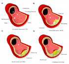
Figure 1
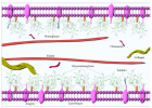
Figure 2
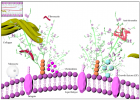
Figure 3

Figure 4
Similar Articles
-
Left Atrial Remodeling is Associated with Left Ventricular Remodeling in Patients with Reperfused Acute Myocardial InfarctionChristodoulos E. Papadopoulos*,Dimitrios G. Zioutas,Panagiotis Charalambidis,Aristi Boulbou,Konstantinos Triantafyllou,Konstantinos Baltoumas,Haralambos I. Karvounis,Vassilios Vassilikos. Left Atrial Remodeling is Associated with Left Ventricular Remodeling in Patients with Reperfused Acute Myocardial Infarction. . 2016 doi: 10.29328/journal.jccm.1001001; 1: 001-008
-
Mid-Ventricular Ballooning in Atherosclerotic and Non-Atherosclerotic Abnormalities of the Left Anterior Descending Coronary ArteryStefan Peters*. Mid-Ventricular Ballooning in Atherosclerotic and Non-Atherosclerotic Abnormalities of the Left Anterior Descending Coronary Artery. . 2016 doi: 10.29328/journal.jccm.1001002; 1:
-
Concentration Polarization of Ox-LDL and Its Effect on Cell Proliferation and Apoptosis in Human Endothelial CellsShijie Liu*,Jawahar L Mehta,Yubo Fan,Xiaoyan Deng,Zufeng Ding*. Concentration Polarization of Ox-LDL and Its Effect on Cell Proliferation and Apoptosis in Human Endothelial Cells. . 2016 doi: 10.29328/journal.jccm.1001003; 1:
-
Intermittent Left Bundle Branch Block: What is the Mechanism?Hussam Ali*,Riccardo Cappato. Intermittent Left Bundle Branch Block: What is the Mechanism?. . 2017 doi: 10.29328/journal.jccm.1001004; 2:
-
Congenital Quadricuspid Aortic Valve, a Rare Cause of Aortic Insufficiency in Adults: Case ReportCyrus Kocherla*,Kalgi Modi. Congenital Quadricuspid Aortic Valve, a Rare Cause of Aortic Insufficiency in Adults: Case Report. . 2017 doi: 10.29328/journal.jccm.1001005; 2: 003-007
-
Short and Medium-Term Evaluation of Patients in Coronary Post-Angioplasty: Préliminary results at the Cardiology Department of the Hospital University Aristide Le Dantec of Dakar (Senegal): Study on 38 CasesDioum M*,Aw F,Masmoudi K,Gaye ND,Sarr SA,Ndao SCT, Mingou J,Ngaidé AA,Diack B,Bodian M,Ndiaye MB,Diao M,Ba SA. Short and Medium-Term Evaluation of Patients in Coronary Post-Angioplasty: Préliminary results at the Cardiology Department of the Hospital University Aristide Le Dantec of Dakar (Senegal): Study on 38 Cases. . 2017 doi: 10.29328/journal.jccm.1001006; 2: 008-012
-
Indications and Results of Coronarography in Senegalese Diabetic Patients: About 45 CasesNdao SCT*,Gaye ND,Dioum M,Ngaide AA,Mingou JS,Ndiaye MB, Diao M,Ba SA. Indications and Results of Coronarography in Senegalese Diabetic Patients: About 45 Cases. . 2017 doi: 10.29328/journal.jccm.1001007; 2: 013-019
-
Procedure utilization, latency and mortality: Weekend versus Weekday admission for Myocardial InfarctionNader Makki,David M Kline,Arun Kanmanthareddy,Hansie Mathelier,Satya Shreenivas,Scott M Lilly*. Procedure utilization, latency and mortality: Weekend versus Weekday admission for Myocardial Infarction. . 2017 doi: 10.29328/journal.jccm.1001008; 2: 020-025
-
Spontaneous rupture of a giant Coronary Artery Aneurysm after acute Myocardial InfarctionOğuzhan Çelik,Mucahit Yetim,Tolga Doğan,Lütfü Bekar,Macit Kalçık*,Yusuf Karavelioğlu. Spontaneous rupture of a giant Coronary Artery Aneurysm after acute Myocardial Infarction. . 2017 doi: 10.29328/journal.jccm.1001009; 2: 026-028
-
Thrombolysis, the only Optimally Rapid Reperfusion TreatmentVictor Gurewich*. Thrombolysis, the only Optimally Rapid Reperfusion Treatment. . 2017 doi: 10.29328/journal.jccm.1001010; 2: 029-034
Recently Viewed
-
Overview on liquid chromatography and its greener chemistry applicationAdel E Ibrahim,Magda Elhenawee,Hanaa Saleh,Mahmoud M Sebaiy*. Overview on liquid chromatography and its greener chemistry application. Ann Adv Chem. 2021: doi: 10.29328/journal.aac.1001023; 5: 004-012
-
COPD and low plasma vitamin D levels: Correlation or causality?Luca Gallelli*, Erika Cione,Stefania Zampogna, Gino Scalone. COPD and low plasma vitamin D levels: Correlation or causality? . J Pulmonol Respir Res. 2018: doi: 10.29328/journal.jprr.1001008; 2: 011-012
-
Environmental Factors Affecting the Concentration of DNA in Blood and Saliva Stains: A ReviewDivya Khorwal*, GK Mathur, Umema Ahmed, SS Daga. Environmental Factors Affecting the Concentration of DNA in Blood and Saliva Stains: A Review. J Forensic Sci Res. 2024: doi: 10.29328/journal.jfsr.1001057; 8: 009-015
-
Markov Chains of Molecular Processes of Biochemical MaterialsOrchidea Maria Lecian*. Markov Chains of Molecular Processes of Biochemical Materials. Int J Phys Res Appl. 2024: doi: 10.29328/journal.ijpra.1001076; 7: 001-005
-
Generation of Curved Spacetime in Quantum FieldSarfraj Khan*. Generation of Curved Spacetime in Quantum Field. Int J Phys Res Appl. 2024: doi: 10.29328/journal.ijpra.1001077; 7: 006-009
Most Viewed
-
Evaluation of Biostimulants Based on Recovered Protein Hydrolysates from Animal By-products as Plant Growth EnhancersH Pérez-Aguilar*, M Lacruz-Asaro, F Arán-Ais. Evaluation of Biostimulants Based on Recovered Protein Hydrolysates from Animal By-products as Plant Growth Enhancers. J Plant Sci Phytopathol. 2023 doi: 10.29328/journal.jpsp.1001104; 7: 042-047
-
Sinonasal Myxoma Extending into the Orbit in a 4-Year Old: A Case PresentationJulian A Purrinos*, Ramzi Younis. Sinonasal Myxoma Extending into the Orbit in a 4-Year Old: A Case Presentation. Arch Case Rep. 2024 doi: 10.29328/journal.acr.1001099; 8: 075-077
-
Feasibility study of magnetic sensing for detecting single-neuron action potentialsDenis Tonini,Kai Wu,Renata Saha,Jian-Ping Wang*. Feasibility study of magnetic sensing for detecting single-neuron action potentials. Ann Biomed Sci Eng. 2022 doi: 10.29328/journal.abse.1001018; 6: 019-029
-
Pediatric Dysgerminoma: Unveiling a Rare Ovarian TumorFaten Limaiem*, Khalil Saffar, Ahmed Halouani. Pediatric Dysgerminoma: Unveiling a Rare Ovarian Tumor. Arch Case Rep. 2024 doi: 10.29328/journal.acr.1001087; 8: 010-013
-
Physical activity can change the physiological and psychological circumstances during COVID-19 pandemic: A narrative reviewKhashayar Maroufi*. Physical activity can change the physiological and psychological circumstances during COVID-19 pandemic: A narrative review. J Sports Med Ther. 2021 doi: 10.29328/journal.jsmt.1001051; 6: 001-007

HSPI: We're glad you're here. Please click "create a new Query" if you are a new visitor to our website and need further information from us.
If you are already a member of our network and need to keep track of any developments regarding a question you have already submitted, click "take me to my Query."


