Abstract
Research Article
Clinical relevance linked to echocardiography diagnosis in Bland, White and Garland syndrome
Mariela Céspedes Almira*, Adel Eladio González Morejón, Giselle Serrano Ricardo and Tania Rosa González Rodríguez
Published: 06 March, 2020 | Volume 5 - Issue 1 | Pages: 051-055
Introduction: Bland, White and Garland syndrome is a coronary anomaly with high mortality without treatment. Its clinical presentation is varied which makes epidemiological documentation difficult. Echocardiography is a useful non-invasive tool for diagnosis.
Objective: To determine the echocardiographic variables that lead to the diagnosis of Bland, White and Garland syndrome and their clinical relevance.
Material: Observational, prospective and cross-sectional study in 31 patients of the “William Soler” Pediatric Cardiocenter, from 2005 to 2018. To check the association of echocardiographic variables with the diagnosis of Bland, White and Garland syndrome, an effectiveness study was carried out that included the analysis of the incidence of echocardiographic variables that lead to the diagnosis of this entity. The clinical relevance was estimated according to the minimum importance limit. The statistical validation of the research results adopted a significance level of less than 5% (p < 0.05).
Results: The variables that facilitate the echocardiographic diagnosis of Bland, White and Garland syndrome were the echocardiographic visualization of the anomalous connection and the reversed flow in the anomalous left coronary artery. These echocardiographic measures have clinical relevance according to the quantification of risk estimators (incidence) the echocardiographic visualization of the anomalous connection, RR 39.00 and the reversed flow in the anomalous coronary artery, RR 26.31. LIM´s calculation value amounted to 6.31 and coincided with the risk estimators (incidence).
Conclusion: The echocardiographic visualization of the anomalous origin of the left coronary artery from the pulmonary arterial trunk and the detection of the local intracoronary reversed flow instituted as factors to be considered for the effective diagnosis of the disease. The documentation of the diagnostic aspects of the syndrome through echocardiography contains high statistical value and clinical relevance.
Read Full Article HTML DOI: 10.29328/journal.jccm.1001086 Cite this Article Read Full Article PDF
Keywords:
Bland; White y Garland syndrome; Clinical relevance; Incidence
References
- Ramírez RF, Bitar HP, Paolinelli GP, Pérez CD, Furnaro F. Anomalías congénitas de arterias coronarias, estudio de aquellas con importancia hemodinámica. Rev Chil Radiol. 2018; 24: 142-50.
- López LP, Centella Hernández T, López Menéndez J, Cuerpo Caballero G, Silva Guisasola J, et al. Registro de intervenciones en pacientes con cardiopatía congénita de la Sociedad Española de Cirugía Torácica-Cardiovascular 2017 y retrospectiva de los últimos 6 años. Cirug Cardiov. 2019; 26: 28-38.
- Perloff JK. Anomalous origin of the left coronary artery from the pulmonary trunk. In the clinical recognition of congenital heart disease. 4th edition. Perloff JK, editor. Philadelphia: WB Saunders Co. 1994; 546-561.
- Wen-xiu L, Bin G, Jiang W, et al. The echocardiografphy diagnosis of infantile and adult types of anomalous origin of the left coronary artery from the pulmonary artery. Chin J Evid Based Pediatr. 2014; 9: 186-189.
- Arunamata A, Buccola Stauffer KJ, Punn R, Chan FP, Maeda K, et al. Diagnosis of anomalous aortic origin of the left coronary artery in a pediatric patient. World J Pediatr Congenit Heart Surg. 2015; 6: 470-473. PubMed: https://www.ncbi.nlm.nih.gov/pubmed/26180168
- Yarrabolu TR, Ozcelik N, Quinones J, Brown MD, Balaguru D. Anomalous origin of left coronary artery from pulmonary artery-duped by 2D; saved by color Doppler: echocardiographic lesson from two cases. Ann Pediatr Cardiol. 2014; 7: 230-232. PubMed: https://www.ncbi.nlm.nih.gov/pmc/articles/PMC4189245/
- Mertens LL, Rigby ML, Horowitz ES, et al. Cross sectional echocardiographic and doppler imaging. In: Anderson RH, editor. Paediatric Cardiology. Philadelphia: Churchill Livingstone Elsevier Ltd; 2010; 313 – 339.
- Lang RM, Badano LP, Mor-Avi V, Afilalo J, Armstrong A, et al. Recommendations for cardiac chamber quantification by echocardiography in adults: an update from the American Society of Echocardiography and the European Association of Cardiovascular Imaging. J Am Soc Echocardiogr. 2015; 28: 1-39. PubMed: https://www.ncbi.nlm.nih.gov/pubmed/25559473
- Cordero L, Rodríguez J, Zuluaga J, Mendoza F, Pérez O. Utilidad de la ecocardiografía en la detección de la insuficiencia cardíaca en un adulto joven con síndrome de origen anómalo de la arteria coronaria izquierda del tronco de la arteria pulmonar y válvula mitral asimétrica similar al paracaídas. Rev Colomb Card. 2018; 25:151.
- Weigand J, Marshall CD, Bacha EA, Chen JM, Richmond ME. Repair of anomalous left coronary artery from the pulmonary artery in the modern era: preoperative predictors of immediate postoperative outcomes and long term cardiac follow‑ Pediatr Cardiol. 2015; 36: 489-497. PubMed: https://www.ncbi.nlm.nih.gov/pubmed/25301273
- Holst LM, Helvind M, Andersen HO. Diagnosis and prognosis of anomalous origin of the left coronary artery from the pulmonary artery. Dan Med J. 2015; 62: A5125. PubMed: https://www.ncbi.nlm.nih.gov/pubmed/26324080
- Sackett DL, Rosenberg WM, Gray JA. Evidence based medicine: what it is and what it is not. BMJ. 1996; 312: 71-2. PubMed: https://www.ncbi.nlm.nih.gov/pubmed/8555924
- García Garmendia JL, Maroto Monserrat F. Interpretación de resultados estadísticos. Med Int. 2018; 42: 370-379.
- Landa Ramírez E, Martínez Basurto AE, Sánchez Sosa JJ. Medicina basada en la evidencia y su importancia en la medicina conductual. Psicología y Salud. 2013; 23: 273-282.
- Laporte JR. Principios básicos de investigación clínica. 2da. ed. Barcelona: Astra Zeneca; 2001.
- Tajer CD. Ensayos terapéuticos, significación estadística y relevancia clínica. Rev Argent Cardiol. 2010; 78: 385-390.
- Bonmatí AN, Vasallo JM. Estadística básica en Ciencias de la Salud. Univ Alicante. 2016:7-59.
- Moncho J. Estadística aplicada a las Ciencias de la Salud. Barcelona: Elsevier; 2015; 62-92.
- Ochoa Sangrador C. Evaluación de la importancia de los resultados de estudios clínicos. Importancia clínica frente a significación estadística. Evid Pediatr. 2010; 6: 40-50.
- Bilder, Christopher R. Loughin, Thomas M. Analysis of Categorical Data with R. First ed. USA: Chapman and Hall/CRC; 2014; 378-382.
- World Medical Association (WMA). World Medical Association Inc. Declaration of Helsinki-Ethical Principles for Medical Research Involving Human Subjects [homepage en Internet]; 64ª Asamblea General, Fortaleza, Brasil, octubre 2013 [citado 16 de marzo de 2019]. Disponible en: https://www.wma.net/en/30publications/10policies/b3/index.html
- Cañedo Andalia R. Medicina basada en la evidencia: un nuevo reto al profesional de la información en salud. ACIMED [serie en Internet]. 2001 [citado 16 de marzo de 2019; 9. Disponible en: http://bvs.sld.cu/revistas/aci/vol9_1_01/aci011001.htm
- Alva Díaz C, Aguirre Quispe W, Becerra Becerra Y, García Mostajo J, Huerta Rosario M, et al. La medicina científica y el programa medicina basada en evidencia han fracasado? Educ Med. 2018; 19: 198-202.
- Baptista González HA. El número necesario a tratar (NNT) y número necesario para hacer daño (NNH). Valoración de la magnitud de la relación beneficio vs. riesgo en las intervenciones médicas. Rev Invest Med Sur Mex. 2008; 15: 302-305.
- Silva Fuente-Alba C, Molina Villagra M. Likelihood ratio (razón de verosimilitud): definición y aplicación en Radiología. Rev Argent Radiol. 2017; 81: 204-208.
- Straus, Glasziou, Richardson, Haynes. Medicina basada en la evidencia: Cómo practicar y enseñar la medicina basada en la evidencia. 5e ed. España: Editorial Elsevier; 2019.
- Ortega Páez E. Sigue vigente hoy día la medicina basada en la evidencia? Rev Pediatr Aten Primaria. 2018; 20: 323-328.
Figures:
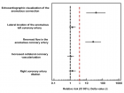
Figure 1
Similar Articles
-
Incidence of symptom-driven Coronary Angiographic procedures post-drug-eluting Balloon treatment of Coronary Artery drug-eluting stent in-stent Restenosis-does it matter?Victor Voon*,Dikshaini Gumani,Calvin Craig,Ciara Cahill,Khalid Mustafa,Terry Hennessy,Samer Arnous,Thomas Kiernan. Incidence of symptom-driven Coronary Angiographic procedures post-drug-eluting Balloon treatment of Coronary Artery drug-eluting stent in-stent Restenosis-does it matter?. . 2017 doi: 10.29328/journal.jccm.1001011; 2: 035-041
-
Electrocardiographic criteria in founder mutations related to Arrhythmogenic cardiomyopathyStefan Peters*. Electrocardiographic criteria in founder mutations related to Arrhythmogenic cardiomyopathy. . 2018 doi: 10.29328/journal.jccm.1001021; 3: 006-007
-
Assessment of risk factors and MACE rate among occluded and non-occluded NSTEMI patients undergoing coronary artery angiography: A retrospective cross-sectional study in Multan, PakistanIbtasam Ahmad,Muhammad Haris,Amnah Javed,Muhammad Azhar*. Assessment of risk factors and MACE rate among occluded and non-occluded NSTEMI patients undergoing coronary artery angiography: A retrospective cross-sectional study in Multan, Pakistan. . 2018 doi: 10.29328/journal.jccm.1001023; 3: 023-030
-
Is there an ideal blood pressure during cardiopulmonary bypass to prevent postoperative cerebral injury? – What does the recent evidence say?Ahmed Zaky*. Is there an ideal blood pressure during cardiopulmonary bypass to prevent postoperative cerebral injury? – What does the recent evidence say?. . 2018 doi: 10.29328/journal.jccm.1001031; 3: 104-105
-
Impact of the Israeli attacks at 2014 on incidence of STEMI in GazaMohammed Habib*,Belal Aldabbour. Impact of the Israeli attacks at 2014 on incidence of STEMI in Gaza. . 2019 doi: 10.29328/journal.jccm.1001037; 4: 036-037
-
C-reactive protein is associated with ventricular repolarization dispersion among patients with metabolic syndromeYlber Jani*,Atila Rexhepi,Bekim Pocesta,Ahmet Kamberi,Fatmir Ferati,Sotiraq Xhunga, Artur Serani,Dali Lala,Agim Zeqiri,Arben Mirto. C-reactive protein is associated with ventricular repolarization dispersion among patients with metabolic syndrome. . 2019 doi: 10.29328/journal.jccm.1001040; 4: 043-052
-
Preclinical stiff heart is a marker of cardiovascular morbimortality in apparently healthy populationCharles Fauvel,Michael Bubenheim,Olivier Raitière,Charlotte Vallet,Nassima Si Belkacem,Fabrice Bauer*. Preclinical stiff heart is a marker of cardiovascular morbimortality in apparently healthy population. . 2019 doi: 10.29328/journal.jccm.1001045; 4: 083-089
-
Do beta adrenoceptor blocking agents provide the same degree of clinically convincing morbidity and mortality benefits in patients with chronic heart failure? A literature reviewMartin Mumuni Danaah Malick*. Do beta adrenoceptor blocking agents provide the same degree of clinically convincing morbidity and mortality benefits in patients with chronic heart failure? A literature review. . 2019 doi: 10.29328/journal.jccm.1001063; 4: 182-186
-
A Systematic review for sudden cardiac death in hypertrophic cardiomyopathy patients with Myocardial Fibrosis: A CMR LGE StudySahadev T Reddy,Antonio T Paladino,Nackle J Silva,Mark Doyle,Diane A Vido,Robert WW Biederman*. A Systematic review for sudden cardiac death in hypertrophic cardiomyopathy patients with Myocardial Fibrosis: A CMR LGE Study. . 2019 doi: 10.29328/journal.jccm.1001064; 4: 187-191
-
Left ventricular ejection fraction and contrast induced acute kidney injury in patients undergoing cardiac catheterization: Results of retrospective chart reviewFiras Ajam*,Obiora Maludum,Nene Ugoeke,Hetavi Mahida,Anas Alrefaee,Amy Quinlan DNP,Jennifer Heck-Kanellidis NP,Dawn Calderon DO,Mohammad A Hossain*,Arif Asif. Left ventricular ejection fraction and contrast induced acute kidney injury in patients undergoing cardiac catheterization: Results of retrospective chart review. . 2019 doi: 10.29328/journal.jccm.1001066; 4: 195-198
Recently Viewed
-
Response of Chemical Fertilisation on Six-year-old Oil Palm Production in Shambillo-Padre Abad- UcayaliEfraín David Esteban Nolberto, Guillermo Gomer Cotrina Cabello*, Robert Rafael-Rutte, Jorge Luis Bringas Salvador, Mag. Carmen Luisa Aquije Dapozzo, Guillermo Vilchez Ochoa, Luis Alfredo Zúñiga Fiestas, Nancy Ochoa Sotomayor, Mg. Merici Medina Guerre. Response of Chemical Fertilisation on Six-year-old Oil Palm Production in Shambillo-Padre Abad- Ucayali. Arch Food Nutr Sci. 2024: doi: 10.29328/journal.afns.1001058; 8: 024-028
-
Forensic analysis of private browsing mechanisms: Tracing internet activitiesHasan Fayyad-Kazan*,Sondos Kassem-Moussa,Hussin J Hejase,Ale J Hejase. Forensic analysis of private browsing mechanisms: Tracing internet activities. J Forensic Sci Res. 2021: doi: 10.29328/journal.jfsr.1001022; 5: 012-019
-
En Bloc Palmar Desquamation in Extensive ChickenpoxSudipta Mondal*. En Bloc Palmar Desquamation in Extensive Chickenpox. Arch Case Rep. 2024: doi: 10.29328/journal.acr.1001100; 8: 078-078
-
Ciliated Hepatic Cyst: Report of a Case and Review of the LiteratureJaime Mejías-Bielsa, Beatriz Agredano-Ávila, Eva Vázquez Tarrio, Ana Teijo-Quintáns*. Ciliated Hepatic Cyst: Report of a Case and Review of the Literature. Arch Case Rep. 2024: doi: 10.29328/journal.acr.1001101; 8: 079-083
-
Advancements in Clinical Research: Phases, Ethical Considerations, and Technological InnovationsAshish Pandey*. Advancements in Clinical Research: Phases, Ethical Considerations, and Technological Innovations. Arch Case Rep. 2024: doi: 10.29328/journal.acr.1001102; 8: 084-086
Most Viewed
-
Evaluation of Biostimulants Based on Recovered Protein Hydrolysates from Animal By-products as Plant Growth EnhancersH Pérez-Aguilar*, M Lacruz-Asaro, F Arán-Ais. Evaluation of Biostimulants Based on Recovered Protein Hydrolysates from Animal By-products as Plant Growth Enhancers. J Plant Sci Phytopathol. 2023 doi: 10.29328/journal.jpsp.1001104; 7: 042-047
-
Sinonasal Myxoma Extending into the Orbit in a 4-Year Old: A Case PresentationJulian A Purrinos*, Ramzi Younis. Sinonasal Myxoma Extending into the Orbit in a 4-Year Old: A Case Presentation. Arch Case Rep. 2024 doi: 10.29328/journal.acr.1001099; 8: 075-077
-
Feasibility study of magnetic sensing for detecting single-neuron action potentialsDenis Tonini,Kai Wu,Renata Saha,Jian-Ping Wang*. Feasibility study of magnetic sensing for detecting single-neuron action potentials. Ann Biomed Sci Eng. 2022 doi: 10.29328/journal.abse.1001018; 6: 019-029
-
Pediatric Dysgerminoma: Unveiling a Rare Ovarian TumorFaten Limaiem*, Khalil Saffar, Ahmed Halouani. Pediatric Dysgerminoma: Unveiling a Rare Ovarian Tumor. Arch Case Rep. 2024 doi: 10.29328/journal.acr.1001087; 8: 010-013
-
Physical activity can change the physiological and psychological circumstances during COVID-19 pandemic: A narrative reviewKhashayar Maroufi*. Physical activity can change the physiological and psychological circumstances during COVID-19 pandemic: A narrative review. J Sports Med Ther. 2021 doi: 10.29328/journal.jsmt.1001051; 6: 001-007

HSPI: We're glad you're here. Please click "create a new Query" if you are a new visitor to our website and need further information from us.
If you are already a member of our network and need to keep track of any developments regarding a question you have already submitted, click "take me to my Query."


